What are the other Names for this Condition? (Also known as/Synonyms)
- Retinal Pigment Epithelial Detachment
- Retinal Tear
- Separation of Retinal Layers
What is Retinal Detachment? (Definition/Background Information)
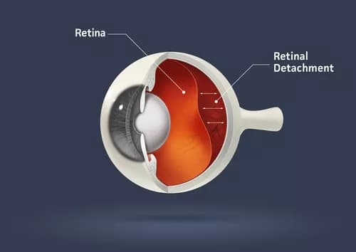
- Retina is a light-sensitive layer of tissue at the back of the eye, which helps to see images focused on it by the cornea and lens
- Retinal Detachment is an eye disorder, wherein the retina gets separated from the underlying layer of blood vessels, which supplies oxygen and other nutrients to it. The condition is a medical emergency
- When the retina gets detached, the supply of oxygen and nutrients are stopped. The onset of symptoms, such as eye floaters and blurred vision, is often sudden. If the condition is left untreated, it may even lead to a complete vision loss and blindness
Different types of Retinal Detachment (classified as per the mechanism of its occurrence) include:
- Rhegmatogenous Retinal Detachment (RRD): It is a sight-threatening eye condition characterized by the separation of the retina from the underlying tissue due to the presence of a retinal tear or hole, allowing vitreous fluid to enter the subretinal space. This type of detachment often requires surgical repair to close the tear and reattach the retina
- Tractional Retinal Detachment (TRD): It occurs when the retina is pulled away from its normal position by fibrous or scar tissue that forms on the retinal surface. Conditions like diabetic retinopathy or proliferative vitreoretinopathy can lead to TRD. Surgical intervention may be needed to relieve the traction forces and reposition the retina
- Exudative Retinal Detachment (ERD): It is characterized by the accumulation of fluid in the layers of the retina, causing it to detach. Unlike other forms of detachment, ERD is often due to factors like vascular abnormalities, inflammation, or tumors, rather than retinal tears. Treatment focuses on addressing the underlying cause of fluid leakage to prevent further detachment and preserve vision
Who gets Retinal Detachment? (Age and Sex Distribution)
- Any individual is susceptible to Retinal Detachment, but the condition is generally observed in individuals in the age group of 40-70 years
- Those who have undergone cataract surgery and those with high myopia, are at a higher risk of contracting this eye disorder
- Worldwide, individuals of both male and female genders and all racial groups and ethnicities are susceptible to this condition
What are the Risk Factors for Retinal Detachment? (Predisposing Factors)
The factors that increase the risk of Retinal Detachment incidence include:
- Adults above the age of 40 years
- A previous occurrence of Retinal Detachment in any of the eyes
- A family history of the condition
- Individuals with severe nearsightedness/myopia (greater than 5 or 6 Diopters) termed degenerative myopia
- Any previous eye surgery, such as for cataract removal or glaucoma
- Congenital abnormalities affecting the eye
- Any past trauma or eye injury (including head injury)
- Use of certain eye drops, such as pilocarpine, for treatment of glaucoma - a condition of elevated eye pressure
- Certain disorders affecting the eye such as chronic inflammation of the eye (known as uveitis), retinoschisis, or diabetic retinopathy
- Participation in certain rough/contact sports such as boxing
It is important to note that having a risk factor does not mean that one will get the condition. A risk factor increases one's chances of getting a condition compared to an individual without the risk factors. Some risk factors are more important than others.
Also, not having a risk factor does not mean that an individual will not get the condition. It is always important to discuss the effect of risk factors with your healthcare provider.
What are the Causes of Retinal Detachment? (Etiology)
- A Detached Retina mostly occurs as a result of a retinal break, hole, or tear. This happens when the vitreous fluid (gel-like material that fills the inside of the eye) contracts, sticks to the retina, and tugs the retina with enough force, causing it to tear
- When the retina gets torn, liquid from the vitreous gel gets accumulated behind the retina leading to its detachment from the back of the eye
The possible causes for Retinal Detachment include:
- Bending or drooping of the vitreous that can occur with advancing age, due to a change in its consistency
- Advanced stages of diabetes
- Trauma/injury to the eye
- Any eye inflammatory disorder may trigger Retinal Detachment
What are the Signs and Symptoms of Retinal Detachment?
Common signs and symptoms exhibited by Retinal Detachment include:
- Early signs may include:
- Sudden appearance of ‘flashing’ bright lights, especially in the peripheral vision
- Vision becomes blurred
- The individuals experience illusions that some small objects like spots, hairs, or strings ,are moving in the eye (called floaters)
- A gray shadow keeps appearing in the vision field
- Partial blindness in the affected eye
Often, only one eye is affected. However, under certain circumstances, such as cataract surgery in both eyes or certain disorders/trauma that affect both the eyes, Retinal Detachment may be bilateral (involving both eyes).
How is Retinal Detachment Diagnosed?
Various tests performed to diagnose Retinal Detachment are aimed at:
- Checking the pupil and retinal response
- Understanding the individual’s ability to visualize colors
Some of the tests performed include:
- Physical examination and evaluation of complete medical history; examination of the eye following its dilation is important to establish a diagnosis
- Optical coherence tomography (OCT) scan of the eye
- Electroretinogram: It makes a record of all electrical signals produced in the retina, while the individual is using his/her vision (is seeing)
- Measuring the intraocular pressure (the fluid pressure inside the eye)
- Fluorescein angiography: A special eye test that checks the blood flow in the retina and choroid, which are the two layers found at the back of the eye
- Ophthalmoscopy: It is an observation test that is done to view the structures, present at the back of the eye, using an ophthalmoscope
- Refraction test: Also, known as a vision test, it is commonly performed to determine an individual’s prescription for eye glasses/contact lenses
- Retinal photography, which aims to project the photographs of the inner surface of the eye
- Tests to check if the patient has any abnormality in identifying colors
- Visual acuity: An eye test, performed to check the clearness of vision
- Slit-lamp examination: This test uses a special instrument, which gives a 3-dimensional picture of various parts of the eye
- Ultrasound of the eye
Many clinical conditions may have similar signs and symptoms. Your healthcare provider may perform additional tests to rule out other clinical conditions to arrive at a definitive diagnosis.
What are the possible Complications of Retinal Detachment?
The major complication associated with Retinal Detachment is a complete loss of vision (particularly when bilateral vision is affected), which may or may not be restored through surgery.
Additionally, surgical complications of Retinal Detachment may include:
- Need for another surgery
- The retina gets detached again; it did not reattach itself properly
- Bleeding from the eye
- Eye infections
- Development of other eye conditions such as cataract or glaucoma
How is Retinal Detachment Treated?
Most individuals with Retinal Detachment require a surgery, either immediately or within a short time. However, a surgery may not be recommended if there are:
- No symptoms manifested by the condition
- The individual has had a Detached Retina for quite some time
However, in such cases, a further aggravation of the eye condition must be avoided and any related symptoms must be treated appropriately.
When the eye condition is mild, then surgery may be performed at the physician’s clinic, which may include:
- Laser surgery may be performed to close the holes/tears in the retina
- Pneumatic retinopexy: In this procedure, gas bubbles are placed in the eye, to make the retina go back to its original position
When Retinal Detachment is severe, the patient is hospitalized and a surgery performed that may include:
- Use of a scleral buckle, which helps push the eye-wall towards the retina
- Vitrectomy, to remove the gel or scar tissue that keeps pulling/tugging at the retina
In addition to the above, it is important to treat any underlying disorder or injury involving the eye promptly and adequately.
How can Retinal Detachment be Prevented?
The following preventive measures may be adopted to reduce the risk of Retinal Detachment:
- Use of protective eye wear is recommended, when working with hammers, lawn mowers, weed-eaters, fireworks, or any similar device/equipment that can cause an eye injury/damage
- All diabetic individuals are advised to keep their sugar level under control consistently
- Undertake early and appropriate treatment of any eye infection, injury, or disorder
- Consultation with an eye specialist and eye examinations at least once a year is recommended, especially for elderly adults and those who are at risk for a Retinal Detachment
What is the Prognosis of Retinal Detachment? (Outcomes/Resolutions)
The prognosis of Retinal Detachment depends on a set of factors, which include:
- Location of the detachment
- Extent of the detachment
- How early was the treatment begun
- Whether the macula (pigmented spot, near the retina center) is affected. If it is not affected, then generally the prognosis is very good
Most cases of Retinal Detachments can be corrected via surgery; the success rate is reported to be about 90%. However, an early diagnosis and treatment of the condition is very important. However, in some severe and delayed cases, the individual may not be able to completely recover his/her vision.
Additional and Relevant Useful Information for Retinal Detachment:
- Individuals with Retinal Detachment in one eye may later develop a bilateral Retinal Detachment (affecting both the eyes)
- Vitrectomy is a microsurgical procedure that involves removal of the vitreous fluid

Related Articles
Test Your Knowledge
Asked by users
Related Centers
Related Specialties
Related Physicians
Related Procedures
Related Resources
Join DoveHubs
and connect with fellow professionals
0 Comments
Please log in to post a comment.