What are the other Names for this Condition? (Also known as/Synonyms)
- Hobnail Hemangioendothelioma
What is Retiform Hemangioendothelioma? (Definition/Background Information)
- Retiform Hemangioendothelioma is an infrequent, potentially aggressive tumor, involving the blood vessels that branch out like a tree, or a net (retiform)
- They are regarded as a kind of low-grade skin angiosarcoma (a malignant cancer of the blood vessels), since they are sometimes capable of metastasizing
- Retiform Hemangioendothelioma manifests during early adulthood. These tumors are normally located on the skin surface, or lie beneath the skin tissues. It is commonly found on the hands and legs. Infrequently, other sites have been documented, such as the torso and scalp
- They are usually treated by performing a complete surgical removal (of the tumors)
Who gets Retiform Hemangioendothelioma? (Age and Sex Distribution)
- Retiform Hemangioendothelioma is mostly observed in young and middle-aged adults (age between 20-40 years); though, there could be a wider age range
- The tumor is observed to affect both sexes uniformly
- There is no known ethnic/racial preference
What are the Risk Factors for Retiform Hemangioendothelioma? (Predisposing Factors)
The risk factors for Retiform Hemangioendothelioma formation include:
- They seem to be linked to a previously performed radiotherapy (for other cancers). Hence, it may be a radiation-induced tumor
- It is thought to be sometimes associated with a chronic obstruction of the lymph vessels (termed as obstructive lymphedema). Obstructive lymphedema can occur due a variety of reasons including infection, surgical procedures, and congenital defects of lymphatics
- The presence of other skin cancers and human herpes virus 8 infections, have been known to be associated with the tumor development. Studies are being performed to scientifically verify the above mentioned associations
It is important to note that having a risk factor does not mean that one will get the condition. A risk factor increases ones chances of getting a condition compared to an individual without the risk factors. Some risk factors are more important than others.
Also, not having a risk factor does not mean that an individual will not get the condition. It is always important to discuss the effect of risk factors with your healthcare provider.
What are the Causes of Retiform Hemangioendothelioma? (Etiology)
- The exact cause and mechanism of Retiform Hemangioendothelioma formation, is unknown. They are thought to occur spontaneously
- It is suggested that their origin could be related to abnormal blood vessel proliferations, for unknown reasons
- Radiation damage to tissue and infections by human herpes virus 8, have been suggested as a possible cause
What are the Signs and Symptoms of Retiform Hemangioendothelioma?
The presentations are based on the location of Retiform Hemangioendothelioma. The signs and symptoms of the tumor include:
- They usually grow at a slow rate and appear as painful, irregular/nodular, mass lesions. They may also cause reduced motion range and discomfort, when present around a joint
- Superficial tumors might look like birthmarks, appearing as red-blue, violet colored scars
- Most tumors are solitary, which means they occur as a single mass. Rarely, multiple lesions have been observed
- Retiform Hemangioendothelioma tumor most commonly occurs in the lower limbs. The next common site of the tumor is the upper limbs. Some case reports have indicated that the tumor can occur in the torso, pelvis, scalp, and penis
How is Retiform Hemangioendothelioma Diagnosed?
A diagnosis of Retiform Hemangioendothelioma is performed using the following diagnostic methods:
- Physical examination, with evaluation of patient’s medical history
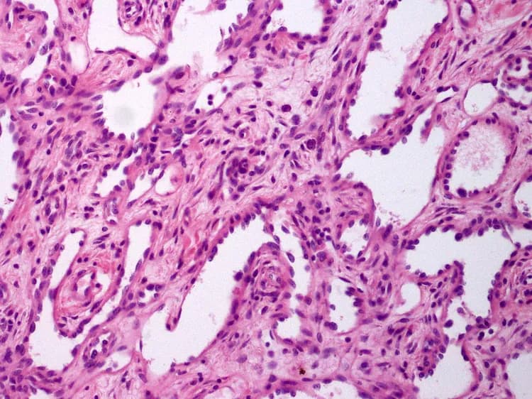
- Histopathological studies conducted on a biopsy specimen, which is examined by a pathologist, who studies the tumor carefully
- MRI scan is performed to determine the extent of the tumor. This MRI study, aids the surgeon by determining the extent of the surgery required
- Differential diagnosis is used to eliminate, other more aggressive types of blood vessel tumors
Many clinical conditions may have similar signs and symptoms. Your healthcare provider may perform additional tests to rule out other clinical conditions to arrive at a definitive diagnosis.
What are the possible Complications of Retiform Hemangioendothelioma?
The complications from Retiform Hemangioendothelioma could include:
- They are known to having a high recurrence rate, after surgical excision
- Metastasizing of the tumor to other body regions; though, this is quite infrequent
- Blood loss during invasive surgical treatment methods may be heavy. Due to this, the blood may accumulate outside the blood vessels, causing a hematoma
- Damage to vital nerves, blood vessels, and surrounding structures during surgery
- Sometimes limb (hand, finger) amputations may have to be performed, while treating Retiform Hemangioendothelioma, in order to achieve a complete surgical excision
How is Retiform Hemangioendothelioma Treated?
Treatment methods for Retiform Hemangioendothelioma include:
- If there are no symptoms, then non-operative measures are adopted (conservative treatment). However, periodic observations are maintained and if there is any pain, it is controlled through pain medication
- Depending on the location, the size of the tumor, and the histological features on the pathology report, any combination of steroids, chemotherapy, radiation therapy, and invasive surgical procedures, are used to treat the tumors
- Interferon injections are used to reduce tumor blood supply, which will help by limiting tumor growth
- Wide surgical excision with removal of the entire lesion is the standard treatment mode used. If the lesion is not fully removed, then the chances of its recurrence are high
- Vascular embolization of the tumor, by blocking the blood vessels feeding the tumor, is used to provide temporary relief from the symptoms, and reduce blood loss during a surgical procedure
- When Retiform Hemangioendothelioma is at an inaccessible location, or is unsafe for surgical intervention, then non-invasive procedures, such as radiation therapy and chemotherapy, are adopted
- Post-operative care is important with minimum activity level to be ensured, until the surgical wound heals. Also, post-operative follow-up care with regular screening and check-ups are important, especially to monitor for any recurrences
How can Retiform Hemangioendothelioma be Prevented?
- Current medical research have not established a way of preventing Retiform Hemangioendothelioma occurrence
- Regular medical screening at periodic intervals with blood tests, radiological scans, and physical examinations, are mandatory for those who have been diagnosed with the tumor
- Due to both its metastasizing potential and high chances of recurrence, often several years of active follow-up, vigilance is necessary
What is the Prognosis of Retiform Hemangioendothelioma? (Outcomes/Resolutions)
- Retiform Hemangioendotheliomas are rare tumors and their behavior is not yet completely understood. However, no fatalities have been reported so far due to this tumor
- The long-term prognosis depends on a combination of factors, such as first appearance/detection of the tumor (duration of the tumor), size and location, its response to surgical treatment and medical therapy, etc.
- Surface and sub-surface tumors that are well-defined can be completely excised, with low chances of recurrence. Deep-seated tumors may be difficult to be completely removed (surgically) and hence, their recurrence rates are higher, compared to superficial tumors
Additional and Relevant Useful Information for Retiform Hemangioendothelioma:
Retiform Hemangioendothelioma is said to be similar to many tumors, but it closely resembles angiosarcoma of the skin (a highly malignant cancer). The pathologist eliminates other tumor types through careful microscopic examination. Eliminating other tumors,during the differential diagnosis, is necessary to arrive at a definitive diagnosis.

Related Articles
Test Your Knowledge
Asked by users
Related Centers
Related Specialties
Related Physicians
Related Procedures
Related Resources
Join DoveHubs
and connect with fellow professionals
0 Comments
Please log in to post a comment.