What are the other Names for this Condition? (Also known as/Synonyms)
- Benign Mixed Tumor of Salivary Gland
- Salivary Gland Pleomorphic Adenoma
What is Pleomorphic Adenoma of Salivary Gland? (Definition/Background Information)
- Pleomorphic Adenoma of Salivary Gland is the most common benign overgrowth arising from the salivary tissue. It may be present in individuals of a wide age range (including children and adults) and normally arises in the parotid gland (major salivary gland)
- The cause of formation of the tumor is unknown, although a few fusion genes have been identified. A past history of radiation therapy to the region is a contributing factor towards Pleomorphic Adenoma of Salivary Gland development
- Most tumors are solitary, small, and asymptomatic. The larger tumors may compress the surrounding structures and cause related signs and symptoms. Sometimes, multiple tumors may be noted that either involve other salivary glands or the same gland
- The diagnosis of Pleomorphic Adenoma of Salivary Gland can be confirmed through a tissue biopsy. The treatment is a complete surgical removal of the tumor with adequate margins
- The prognosis of Pleomorphic Adenoma of Salivary Gland is generally excellent with adequate treatment (complete removal through surgery). However, tumor recurrences are not uncommon, and some are even known to undergo malignant transformations
Who gets Pleomorphic Adenoma of Salivary Gland? (Age and Sex Distribution)
- Between 60-65% of all salivary gland tumors are pleomorphic adenomas; it is the most frequently observed benign salivary gland tumor
- The incidence of Pleomorphic Adenoma of Salivary Gland is about 1 in 35,000 population
- The tumor can be present in individuals of a wide age range, with a predilection for middle-age (mean age of 46 years; age range 30-60 years). It is seen in infants, children, young adults too
- The condition is slightly more common in females, though both males and females are affected
- All races and ethnic groups may be affected
What are the Risk Factors for Pleomorphic Adenoma of Salivary Gland? (Predisposing Factors)
- No clearly identified risk factors for Pleomorphic Adenoma of Salivary Gland have been established to date
- These tumors are seen to arise (or be present) simultaneously with other benign tumors, such as Warthin tumor, in some cases
- A history of radiation therapy to the head and neck region may be a risk factor for the development of this tumor. The radiation treatment may have been undertaken even 15-20 years’ back
It is important to note that having a risk factor does not mean that one will get the condition. A risk factor increases one’s chances of getting a condition compared to an individual without the risk factors. Some risk factors are more important than others.
Also, not having a risk factor does not mean that an individual will not get the condition. It is always important to discuss the effect of risk factors with your healthcare provider.
What are the Causes of Pleomorphic Adenoma of Salivary Gland? (Etiology)
Currently, the cause of formation of Pleomorphic Adenomas of Salivary Gland is unknown.
- However, based on extensive studies, pleomorphic adenomas have shown chromosomal rearrangements, translocations, and other genetic abnormalities
- Genetic mutations involving 2 specific genes (PLAG1 and HMGA2) have been noted:
- The PLAG1 gene on chromosome 8 is known to form fusion with any of the CTNNB1, LIFR, and SII genes
- The HMGA2 gene on chromosome 12 is known to form fusion with either NF1B or FHIT gene
- The above fusion genes are specifically observed with Salivary Gland Pleomorphic Adenomas and can help in making a definite diagnosis
Some studies have also informed about the involvement of the cancer-causing virus SV40 (simian virus 40). It is researched that this virus may provoke the development of Pleomorphic Adenoma of Salivary Gland.
What are the Signs and Symptoms of Pleomorphic Adenoma of Salivary Gland?
The signs and symptoms of Pleomorphic Adenoma of Salivary Gland may include:
- Presence of a tumor mass in the mouth, on the cheek, around the jaw, affecting the soft and hard palate, etc.
- The mass may be smooth, firm, and mobile; most tumors appear to be covered by a mucus-coated capsule (from the inside of the mouth)
- Tumors are typically solitary, but multiple lesions that arise from the same gland or from other salivary glands may be noted
- The tumors are well-defined and spherical or oval
- The size may range from 2-5 cm, although some can grow to very large sizes
- Small tumors usually do not cause any symptoms. Pain is generally not felt, unless the surrounding structures are compressed
- Large tumors may cause skin inflammation and redness
- Most tumors are confined to the major salivary glands; i.e., the parotid glands, in about 80% of the cases. The submandibular glands are affected next (in nearly 10% of the cases)
- Some tumors may occur in the minor salivary glands. If tumors occur within the minor salivary gland of the oral mucosa, it is called an intraoral pleomorphic adenoma
- The remaining tumors can arise at locations, such as the nasal cavity and paranasal sinuses, and even in the food-pipe (pleomorphic adenoma of esophagus) and wind-pipe (pulmonary pleomorphic adenoma)
Large tumors may cause the following signs and symptoms:
- Difficulty in opening one’s mouth; pain while eating (pain may be felt in the mouth and face)
- Numbness of part of the face
- Dryness of mouth
- Inability to move one side of the face due to damage to the facial nerve, known as facial nerve palsy
- Facial pain
- Breathing difficulty
How is Pleomorphic Adenoma of Salivary Gland Diagnosed?
The following exams and procedures may be used in the diagnosis of Pleomorphic Adenoma of Salivary Gland:
- Evaluation of the individual’s medical history and a through physical examination
- Plain X-ray of the head and neck region
- Ultrasound scan of the affected salivary gland
- CT or CAT scan with contrast of the head and neck may show a well-defined mass. This radiological procedure creates detailed 3-dimensional images of structures inside the scanned region
- MRI scans of head and neck region: A magnetic field is used to create high-quality pictures of certain parts of the body, such as tissues, muscles, nerves, and bones. These high-quality pictures may reveal the presence of the tumor
- Fluorescence in situ hybridization (FISH): It is a test performed on the blood or bone marrow cells to detect chromosome changes (cytogenetic analysis). The test helps in identifying genetic abnormalities that may not be evident with an examination of cells under a microscope
- Polymerase chain reaction (PCR): It is used to measure the presence of certain biomarkers in blood or bone marrow cells. The test is ultrasensitive and detects extremely low amounts of biomarkers remaining in blood, which can be missed by cytogenetic methods, such as FISH, karyotype, or flow cytometry. PCR allows a more sensitive follow-up of patients in remission and can help determine whether additional treatment is necessary
Although the above modalities can be used to make an initial diagnosis, a tissue biopsy of the tumor is necessary to make a definitive diagnosis to begin treatment. The tissue for diagnosis can be procured in multiple different ways which include:
- Fine needle aspiration (FNA) biopsy of the tumor: A FNA biopsy may not be helpful, because one may not be able to visualize the different morphological areas of the tumor. Hence, a FNA biopsy as a diagnostic tool has certain limitations, and an open surgical biopsy may be recommended.
- Salivary gland core biopsy of the tumor
- Salivary gland open biopsy of the tumor
Tissue biopsy:
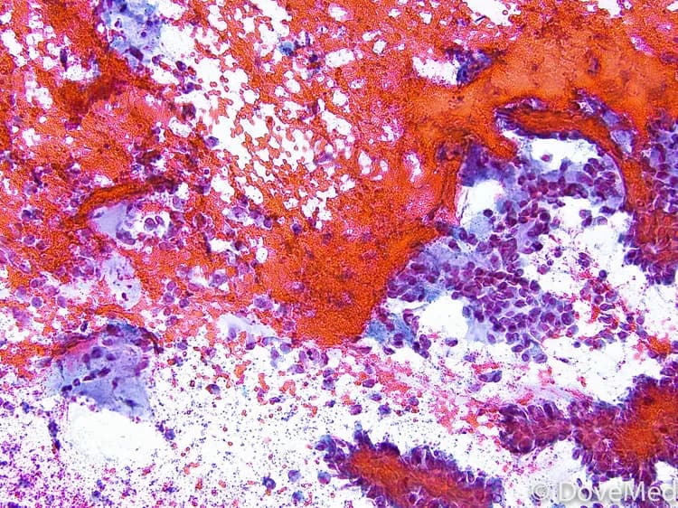
- A tissue biopsy of the tumor is performed and sent to a laboratory for a pathological examination. A pathologist examines the biopsy under a microscope. After putting together clinical findings, special studies on tissues (if needed) and with microscope findings, the pathologist arrives at a definitive diagnosis. Examination of the biopsy under a microscope by a pathologist is considered to be gold standard in arriving at a conclusive diagnosis
- Biopsy specimens are studied initially using Hematoxylin and Eosin staining. The pathologist then decides on additional studies depending on the clinical situation
- Sometimes, the pathologist may perform special studies, which may include immunohistochemical stains, molecular testing, and very rarely, electron microscopic studies to assist in the diagnosis
A differential diagnosis, to eliminate other tumor types are often considered, before arriving at a definitive diagnosis. Pleomorphic Adenoma of Salivary Gland is also known to be misdiagnosed as the following tumors:
- Adenoid cystic carcinoma
- Oncocytoma
Based on the components present in the tumor, on histological examination, the tumor may be termed:
- Cellular pleomorphic adenoma
- Chondroid pleomorphic adenoma (tumor forms cartilage type tissue)
- Hyaline pleomorphic adenoma
- Lipomatous pleomorphic adenoma
- Myxoid pleomorphic adenoma
- Osseous pleomorphic adenoma (tumor with bone formation)
Many clinical conditions may have similar signs and symptoms. Your healthcare provider may perform additional tests to rule out other clinical conditions to arrive at a definitive diagnosis.
What are the possible Complications of Pleomorphic Adenoma of Salivary Gland?
The following complications due to Pleomorphic Adenoma of Salivary Gland may be observed:
- Severe emotional stress and cosmetic concerns (facial disfigurement)
- Large tumors may show the presence of multiple tiny satellite nodules around the main mass
- Even though rare, the tumors are known to recur following surgery to remove them, particularly in younger individuals. According to some studies, the recurrence rate may range from 3-50%. Also, long-term recurrences (even after 10 years) have been observed
- Tumors that recur are known to appear widespread in the oral cavity; they are usually multifocal (arising simultaneously at many locations). The treatment in such cases may be difficult or complicated
- Occasionally, pleomorphic adenomas are known to transform into malignant tumors. Although, this may take place over 10-15 years
- Surgery to remove the tumor mass may result in facial nerve palsy (surgical complication)
- Post-surgical wound infection
How is Pleomorphic Adenoma of Salivary Gland Treated?
- A complete surgical excision with clear or wide margins is the preferred mode of treatment of Pleomorphic Adenoma of Salivary Gland. When tumors involve the parotid gland, a superficial parotidectomy is the treatment of choice
- Tumor recurrence may lead to a need for multiple surgeries and more extensive therapies
- Post-operative care is important: One must maintain minimum activity levels, until the surgical wound heals
- Long-term follow-up is required, because recurrence at the site of surgery have been reported many years (even decades) after surgery
How can Pleomorphic Adenoma of Salivary Gland be Prevented?
- Current medical research has not established a method of preventing the formation of Pleomorphic Adenoma of Salivary Glands
- Regular medical screening at periodic intervals with blood tests, radiological scans, and physical examinations, for those who have already endured the tumor, are helpful
What is the Prognosis of Pleomorphic Adenoma of Salivary Gland? (Outcomes/Resolutions)
- The prognosis of Pleomorphic Adenoma of Salivary Gland is generally excellent with surgical intervention and complete removal, since it is a benign tumor
- However, some tumors are known to recur and recurrent tumors may be disseminated across the oral cavity affecting adjoining structures/regions. It is typically difficult and challenging to remove them in such cases. Based on studies, the rate of recurrence for tumors of the parotid gland is about 3.5% in 5 years, and 7% in 10 years
- Besides recurrence, malignant transformations have been infrequently noted (1 in 10 chance of malignancy). In such cases, the prognosis of Salivary Gland Pleomorphic Adenoma is based on several factors, including on the stage of the tumor
- The factors that may predict a malignant transformation include:
- Advanced age of the patient
- Presence of tumor for a long duration (over 10 years)
- Very large tumor size
- Involvement of the submandibular glands
Additional and Relevant Useful Information for Pleomorphic Adenoma of Salivary Gland:
There are 3 major types of salivary glands and these include the following:
- Parotid glands, found on the sides of the face
- Submandibular glands located at the back of mouth, on both sides of the jaw
- Sublingual glands that are seen under the floor of the mouth
The salivary glands produce saliva that helps in:
- Lubricating the mouth
- Swallowing
- Protects the teeth against bacteria
- Digestion of food


Related Articles
Test Your Knowledge
Asked by users
Related Centers
Related Specialties
Related Physicians
Related Procedures
Related Resources
Join DoveHubs
and connect with fellow professionals
0 Comments
Please log in to post a comment.