What are the other Names for this Condition? (Also known as/Synonyms)
- Ancient Common Mole
- Neurotized Melanocytic Nevus
- Neurotized Naevus
What is Neurotized Nevus? (Definition/Background Information)
- A nevus (plural nevi) is a mole on the skin that can occur on any part of the body. A Neurotized Nevus is a type of mole. In this type, the melanocytes are in the dermis. With time, there is fibrosis around these melanocytes resulting in a benign Neurotized Nevus
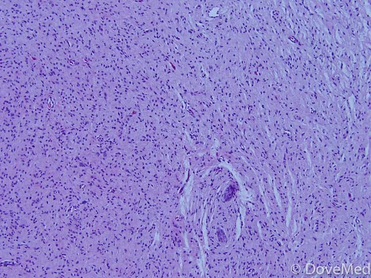
- A diagnosis is not made clinically by examining the patient, since the Neurotized Nevus resembles a common mole. The definitive diagnosis is made by a pathologist under the microscope
- When the nevus is examined by a pathologist under the microscope, the melanocytes appear like spindle cells resembling a nerve; and hence, it is called a Neurotized Nevus
- Normally, Neurotized Nevi are present in all individuals including children and adults. The condition is more common in fair-skinned individuals than dark-skinned individuals. Sun exposure is a risk factor for acquiring the condition
- In many cases, the nevus occurs in a random manner, while few cases are known to run in families. However, the cause of formation of a Neurotized Nevus is generally unknown. Research studies have implicated certain genetic mutations as being the probable cause
- Treatment is generally not required for Neurotized Nevus, unless it presents cosmetic issues. The prognosis is usually excellent with appropriate treatment, since these moles are generally benign
Who gets Neurotized Nevus? (Age and Sex Distribution)
- A Neurotized Nevus may be present at birth or form at any age in an individual. Both children and adults may be observed with this nevus type
- Both males and females are affected and there is no gender bias observed
- All racial and ethnic groups are at risk, but this nevus is more frequent in fair-skinned individuals (Caucasians) than dark-skinned individuals (such as some Africans or Asians)
What are the Risk Factors for Neurotized Nevus? (Predisposing Factors)
The risk factors for Neurotized Nevus may include:
- Sun exposure
- A positive family history of the mole, in some cases
- Generally, lighter-skinned races or individuals (such as Caucasians of America and Europe) are at a higher risk compared to other darker-skinned racial groups (such as Africans and Asians)
It is important to note that having a risk factor does not mean that one will get the condition. A risk factor increases one’s chances of getting a condition compared to an individual without the risk factors. Some risk factors are more important than others.
Also, not having a risk factor does not mean that an individual will not get the condition. It is always important to discuss the effect of risk factors with your healthcare provider.
What are the Causes of Neurotized Nevus? (Etiology)
- The cause of Neurotized Nevus formation is unknown, in a majority of cases. In some cases, certain gene mutations have been documented
- Most cases occur sporadically, meaning in a random fashion (sporadic, non-familial nevus). However, it can run in families too (in which case it is termed a familial nevus)
What are the Signs and Symptoms of Neurotized Nevus?
The signs and symptoms of Neurotized Nevus that may be observed include:
- It is a benign mole of melanocytic cells on the skin. It can occur anywhere on the body, and may be single or many in numbers
- It is usually well defined and less than 10 mm in size. The nevi are usually round or oval; although they can be of any shape
- They can be present as a macule, papule, or nodule (raised mass)
- The benign mole is usually slow-growing and pigmented. The color may vary from skin color to light brown to black. The pigment color over the mole is usually uniform
Neurotized Nevi usually occur early in life and grow, as the child becomes an adult. As one age, these moles may become smaller; in many cases, they are known to disappear, when one reaches the age of about 60 years.
How is Neurotized Nevus Diagnosed?
A Neurotized Nevus is diagnosed through the following tools:
- Complete physical examination with evaluation of medical history
- Dermoscopy: It is a diagnostic tool where a dermatologist examines the skin using a special magnified lens
- Wood’s lamp examination: In this procedure, the healthcare provider examines the skin using ultraviolet light. It is performed to examine the change in skin pigmentation
- Skin biopsy: A skin biopsy is performed and sent to a laboratory for a pathological examination. The pathologist examines the biopsy under a microscope. After putting together clinical findings, special studies on tissues (if needed) and with microscope findings, the pathologist arrives at a definitive diagnosis
Note: In majority of the cases, no biopsy is necessary. But they may be performed to rule out other conditions presenting similar signs and symptoms.
Many clinical conditions may have similar signs and symptoms. Your healthcare provider may perform additional tests to rule out other clinical conditions to arrive at a definitive diagnosis.
What are the possible Complications of Neurotized Nevus?
There are frequently no complications that arise from a Neurotized Nevus.
- Nevertheless, in some individuals, it may give rise to cosmetic concerns
- Scratching or itching of the moles may lead to bleeding and ulceration. This can cause secondary bacterial or fungal infections to develop
How is Neurotized Nevus Treated?
The treatment measures for Neurotized Nevus include:
- The healthcare provider may choose to regularly observe the mole, to check for any atypical features; a “wait and watch” approach may be undertaken
- Surgical excision and removal of the mole, if necessary for cosmetic reasons
- The benign nevus can also be excised through electrocautery surgical procedure
How can Neurotized Nevus be Prevented?
Current medical research has not established a method of preventing Neurotized Nevi. However, the following measures can help prevent these moles from becoming either a dysplastic mole or a melanoma:
- Minimize direct exposure to the sun’s ultraviolet (UV) rays
- Avoid tanning beds and sun lamps
- Perform self-examination of the skin, from head to toe, once a month (especially if you are at a higher risk)
- Get a professional skin exam from a healthcare provider, once a year
What is the Prognosis of Neurotized Nevus? (Outcomes/Resolutions)
- The prognosis of Neurotized Nevus is excellent on its complete excision and removal
- Since these are benign moles, the prognosis is excellent, even if only periodic observation is maintained
Additional and Relevant Useful Information for Neurotized Nevus:
- Common moles/nevi are of 3 types:
- Junctional common mole
- Compound common mole
- Intradermal common mole
- Do not pick or scratch the moles


Related Articles
Test Your Knowledge
Asked by users
Related Centers
Related Specialties
Related Physicians
Related Procedures
Related Resources
Join DoveHubs
and connect with fellow professionals
0 Comments
Please log in to post a comment.