What are the other Names for this Condition? (Also known as/Synonyms)
- Esophageal Leiomyosarcoma
- Leiomyosarcoma of Oesophagus
- Oesophageal Leiomyosarcoma
What is Leiomyosarcoma of Esophagus? (Definition/Background Information)
- Leiomyosarcoma (LMS) is a rare type of connective tissue cancer, accounting for 5-10% of all soft tissue sarcomas (a type of cancer). It was once believed that leiomyosarcomas originated from small, benign, smooth muscle tumors, known as leiomyomas. The occurrence of a malignant tumor from a leiomyoma is now believed to be extremely rare
- Leiomyosarcoma of Esophagus is a very uncommon and highly-malignant mesenchymal (non-epithelial) tumor. The esophagus is a part of the upper gastrointestinal tract and is also known as the ‘food-pipe’
- The cause of formation of Esophageal Leiomyosarcoma may be due to genetic abnormalities. No definitive risk factors are observed for this esophageal tumor, though leiomyosarcomas are known to be associated with trauma to the region, high-dose radiation exposure, and poor immunity
- Small tumors may be painless and asymptomatic, while advanced cases present swallowing difficulties and chest and upper abdominal pain. In case of a delayed diagnosis, metastasis of the leiomyosarcoma to other distant regions may occur
- The treatment of choice is a surgical excision with clear margins followed by radiation therapy/chemotherapy. In case of spread of cancer to other regions, a combination of treatments may be considered by the healthcare provider
- The prognosis depends upon a set of several factors including the stage of the tumor, extent of tumor spread, overall health of the patient, and many other factors. In general, the prognosis of Leiomyosarcoma of Esophagus is poor
Who gets Leiomyosarcoma of Esophagus? (Age and Sex Distribution)
- Leiomyosarcoma of Esophagus is a very rare tumor that is observed in adults
- Both males and females are affected
- All races and ethnic groups are at risk for the tumor
What are the Risk Factors for Leiomyosarcoma of Esophagus? (Predisposing Factors)
The risk factors for Leiomyosarcoma of Esophagus are not well-established. However, there are a few leading theories behind LMS formation:
- Trauma to the affected region
- High-dose radiation exposure is believed to increase the risk for leiomyosarcoma formation
- Certain inherited genetic traits are believed to increase the risk
- Exposure to certain chemical agents, such as vinyl chloride, certain herbicides, and/or dioxins, may increase the risk
- Immunocompromised patients infected by Epstein-Barr virus seem to be predisposed to LMS. The reason for this is not understood, yet there seems to be a definite correlation between the viral infection and the arising of multiple, synchronized leiomyosarcomas
It is important to note that having a risk factor does not mean that one will get the condition. A risk factor increases one’s chances of getting a condition compared to an individual without the risk factors. Some risk factors are more important than others.
Also, not having a risk factor does not mean that an individual will not get the condition. It is always important to discuss the effect of risk factors with your healthcare provider.
What are the Causes of Leiomyosarcoma of Esophagus? (Etiology)
The exact cause of development of Leiomyosarcoma of Esophagus is unknown, but it may be due to certain genetic defects.
- In general, it is known that cancers form when normal, healthy cells begin transforming into abnormal cells - these cancer cells grow and divide uncontrollably (and lose their ability to die), resulting in the formation of a mass or a tumor.
- Many cancer types are caused by genetic mutations. These can occur, due to inherited mutations, or mutations that occur due to environmental factors
- The transformation of normally healthy cells into cancerous cells may be the result of genetic mutations. Mutations allow the cancer cells to grow and multiply uncontrollably to form new cancer cells
- These tumors can invade nearby tissues and adjoining body organs, and even metastasize and spread to other regions of the body
What are the Signs and Symptoms of Leiomyosarcoma of Esophagus?
Leiomyosarcoma of Esophagus may present the following signs and symptoms:
- Initially, small-sized tumors may be asymptomatic
- Most tumor masses appear as polyps (polypoid) on the esophageal wall
- Commonly observed symptoms for advanced cancer are swallowing difficulties (seen in 80-90% of the cases), chest pain (retrosternal pain), upper abdominal pain (epigastric pain), narrowing of esophagus can cause regurgitation or vomiting sensation
- The tumor may be located anywhere in the esophagus
- Esophageal carcinomas spread up and down the food-pipe or around the GI tube
- Large tumors may cause a pressure effect by compressing adjoining structures and organs
- Large tumors may ulcerate and bleed and become painful
- Involvement of lymph nodes may be noted
How is Leiomyosarcoma of Esophagus Diagnosed?
A diagnosis of Leiomyosarcoma of Esophagus may involve the following:
- A thorough medical history and physical examination
- X-ray of the chest
- CT or MRI scan of the chest: For advanced cases and to check cancer growth and spread, including lymph node involvement
- Upper GI endoscopy: An endoscopic procedure is performed using an instrument called an endoscope, which consists of a thin tube and a camera. Using this technique, the radiologist can have a thorough examination of the insides of the upper gastrointestinal tract
- Endoscopic ultrasonography: During this procedure, fine needle aspiration biopsy (FNAB) can be performed on the affected area. This is good technique for tumor detection including tumor invasion parameters, and whether nearby lymph nodes are affected
- Endocytoscopy: It is a non-invasive technique helpful for invasive carcinomas that are located superficially
- Early cancer lesions may be detected using narrow band imaging technique
- Barium swallow
- Whole body PET scans to determine how far the cancer has spread to other organ systems
Although the above modalities can be used to make an initial diagnosis, a tissue biopsy of the tumor is necessary to make a definitive diagnosis to begin treatment. The tissue for diagnosis can be procured in multiple different ways which include:
- Fine needle aspiration (FNA) biopsy of the tumor: A FNA biopsy may not be helpful, because one may not be able to visualize the different morphological areas of the tumor and the tumor may be misdiagnosed. Hence, a FNA biopsy as a diagnostic tool has certain limitations, and an open surgical biopsy is preferred
- Core biopsy of the tumor
- Open biopsy of the tumor
Tissue biopsy:
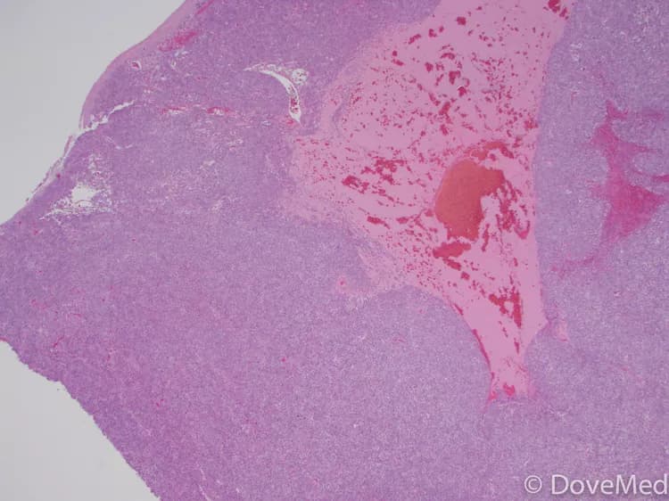
- A tissue biopsy of the tumor is performed and sent to a laboratory for a pathological examination. A pathologist examines the biopsy under a microscope. After putting together clinical findings, special studies on tissues (if needed) and with microscope findings, the pathologist arrives at a definitive diagnosis. Examination of the biopsy under a microscope by a pathologist is considered to be gold standard in arriving at a conclusive diagnosis
- Biopsy specimens are studied initially using Hematoxylin and Eosin staining. The pathologist then decides on additional studies depending on the clinical situation
- Sometimes, the pathologist may perform special studies, which may include immunohistochemical stains, molecular testing, and very rarely, electron microscopic studies to assist in the diagnosis
Many clinical conditions may have similar signs and symptoms. Your healthcare provider may perform additional tests to rule out other clinical conditions to arrive at a definitive diagnosis.
What are the possible Complications of Leiomyosarcoma of Esophagus?
The complications of Leiomyosarcoma of Esophagus may include the following:
- Ulceration of the tumor can lead to secondary infections of bacteria and fungus
- Compression of the underlying nerve, which can affect nerve function
- Severe obstruction of the food-pipe with pain, leading to difficulties in eating. This can lead to extreme weight loss due to malnutrition
- Perforation or rupture of the esophagus can lead to the formation of fistulas, which in turn can cause aspiration pneumonia. This is a life-threatening condition
- Stricture formation of esophagus
- The tumor can metastasize to the local or distant lymph nodes
- Metastasize to other body organs such as the lungs, intestines, abdominal cavity, etc.
- Recurrence of the tumor following treatment
- Side effects of chemotherapy (such as toxicity) and radiation
- Damage to the muscles, vital nerves, and blood vessels, during surgery
- Post-surgical infection at the wound site is a potential complication
How is Leiomyosarcoma of Esophagus Treated?
The treatment of Leiomyosarcoma of Esophagus usually involves surgery, which is the first treatment option considered.
- When the tumor is confined to the surface, then endoscopic mucosal/submucosal resection (or surgical removal via endoscopy) is undertaken
- Esophagectomy or surgery to remove part (or all) of esophagus
- If the tumor has metastasized, then a combination of chemotherapy, radiation therapy, and invasive procedures may be considered; although, the use of chemotherapy and radiation therapy is reportedly controversial
- Palliative care is provided for advanced cancer stages
- Follow-up care with regular screening and check-ups are very important and encouraged
How can Leiomyosarcoma of Esophagus be Prevented?
- Current medical research has not established a method of preventing the formation of Leiomyosarcoma of Esophagus
- Regular medical screening at periodic intervals with blood tests, radiological scans, and physical examinations, are mandatory for those who have been diagnosed with the tumor
- Due to its metastasizing potential and chances of recurrence, often several years of active follow-up and vigilance is recommended
What is the Prognosis of Leiomyosarcoma of Esophagus? (Outcomes/Resolutions)
- The prognosis of Leiomyosarcoma of Esophagus is generally poor. Nevertheless, reports indicate that the prognosis is better than squamous cell carcinomas of the esophagus
- The Kaplan-Meier method indicates that the survival rates for individuals with the tumor is 80% for 3 years, 58% for 5 years, and 31% for 10 years
- The 5 year survival rate for tumors that have infiltrated deep into the tissues is only 25%, while it is 83% for intramural/polypoid tumors
- The prognosis depends upon a set of several factors, which include:
- Stage of tumor: With lower-stage tumors, when the tumor is confined to site of origin, the prognosis is usually excellent with appropriate therapy. In higher-stage tumors, such as tumors with metastasis, the prognosis is poor
- Overall health of the individual
- Age of the individual: Older individuals generally have poorer prognosis than younger individuals
- The size of the tumor: Individuals with small-sized tumors fare better than those with large-sized tumors
- Individuals with bulky disease may have a poorer prognosis
- Involvement of vital organs may complicate the condition
- The surgical resectability of the tumor (meaning, if the tumor can be removed completely)
- Whether the tumor is occurring for the first time, or is a recurrent tumor. Recurring tumors have worse prognosis compared to tumors that do not recur
- Response to treatment: Tumors that respond to treatment have better prognosis compared to tumors that do not respond to treatment; good response to treatment (surgery and/or chemotherapy/radiation therapy)
- Progression of the condition makes the outcome worse
- An early diagnosis and prompt treatment of the tumor generally yields better outcomes than a late diagnosis and delayed treatment
- The combination chemotherapy drugs used, may have some severe side effects (such as cardio-toxicity). This chiefly impacts the elderly adults, or those who are already affected by other medical conditions. Tolerance to the chemotherapy sessions is a positive influencing factor
Additional and Relevant Useful Information for Leiomyosarcoma of Esophagus:
The following DoveMed website links are useful resources for additional information:

Related Articles
Test Your Knowledge
Asked by users
Related Centers
Related Specialties
Related Physicians
Related Procedures
Related Resources
Join DoveHubs
and connect with fellow professionals
0 Comments
Please log in to post a comment.