Cellular Blue Nevus
What are the other Names for this Condition? (Also known as/Synonyms)
- Cellular Blue Mole
- Cellular Blue Naevus
What is Cellular Blue Nevus? (Definition/Background Information)
- A nevus (plural nevi) is a mole on the skin that can occur on any part of the body. A blue nevus is a benign proliferation of melanocytes (which makes melanin pigment) in the dermis or skin that is generally bluish in appearance
- In a Cellular Blue Nevus, the number of melanoma sites is much higher than a regular blue nevus, and hence it is called so. It is an unusual type of mole that in rare cases, can turn into a malignant melanoma
- The mole can be present anywhere on the body, though most of them are seen on the buttocks and lower back. They appear as papules or nodules
- Normally, these moles are present during childhood and adolescence. Cellular Blue Nevi are generally asymptomatic in nature, but may present cosmetic concerns in some individuals
- Sun exposure may be a risk factor for acquiring the condition. Cellular Blue Nevus is observed to occur in a random manner and the cause is unknown
- Once a Cellular Blue Nevus is confirmed through a diagnostic biopsy, it is surgically removed completely, since there is a small probability of the nevus turning into a malignant melanoma. If the diagnostic biopsy completely removed it at the first surgery, no further procedure is generally needed
- The prognosis is generally excellent with or without treatment since these moles are typically benign. However, in case of a malignant transformation, the prognosis depends upon various factors, chiefly the stage of the tumor
Who gets Cellular Blue Nevus? (Age and Sex Distribution)
- Cellular Blue Nevus is a skin condition that may be present at birth or form at any age in an individual. However, children and adolescents are generally observed to have this type of nevus. It is uncommon in older age
- Both males and females are affected and there is no gender bias seen
- All racial and ethnic groups are at risk, but this nevus type is more frequent in fair-skinned individuals (Caucasians)
What are the Risk Factors for Cellular Blue Nevus? (Predisposing Factors)
The risk factors identified for Cellular Blue Nevus include:
- Sun exposure
- Generally, lighter-skinned races or individuals (such as Caucasians of America and Europe) are at a higher risk compared to other darker-skinned racial groups (such as Africans and Asians)
It is important to note that having a risk factor does not mean that one will get the condition. A risk factor increases ones chances of getting a condition compared to an individual without the risk factors. Some risk factors are more important than others.
Also, not having a risk factor does not mean that an individual will not get the condition. It is always important to discuss the effect of risk factors with your healthcare provider.
What are the Causes of Cellular Blue Nevus? (Etiology)
- The cause of Cellular Blue Nevus formation is presently unknown
- It is believed to be acquired due to certain factors that are yet to be identified. The cause may be probably genetic
What are the Signs and Symptoms of Cellular Blue Nevus?
Cellular Blue Nevus may not present any signs and symptoms in most cases. The general features of the condition include:
- It is a benign tumor of melanocytic cells on the skin. They may be single or many in numbers
- It is usually well-defined and ranges from 1-3 cm in size (larger than a common blue nevus). These moles/nevi are usually round or oval in shape; although, they can be of any shape
- They can be present as a papule or nodule (a raised mass). A papule is an area of abnormal skin tissue that is less than 1 centimeter around. Usually a papule has distinct borders, and it can appear in a variety of shapes
- The benign nevus is usually slow-growing and pigmented. The color may vary from blue to blue black
- The pigment color over the mole is usually uniform, although sometimes irregular pigmentation is observed. This can cause concerns of a melanoma requiring further investigations by a healthcare provider
- Cellular Blue Nevi can occur anywhere on the body. However, many of them are seen on the buttocks, lower back, and head and neck region
They usually occur early in life and grow in size, as the child becomes an adult. As one age, these moles may become smaller and in many cases, even disappear.
How is Cellular Blue Nevus Diagnosed?
A Cellular Blue Mole/Nevus is diagnosed through the following tools:
- Complete physical examination with evaluation of medical history
- Dermoscopy: It is a diagnostic tool where a dermatologist examines the skin using a special magnified lens
- Wood’s lamp examination: In this procedure, the healthcare provider examines the skin using ultraviolet light. It is performed to examine the change in skin pigmentation
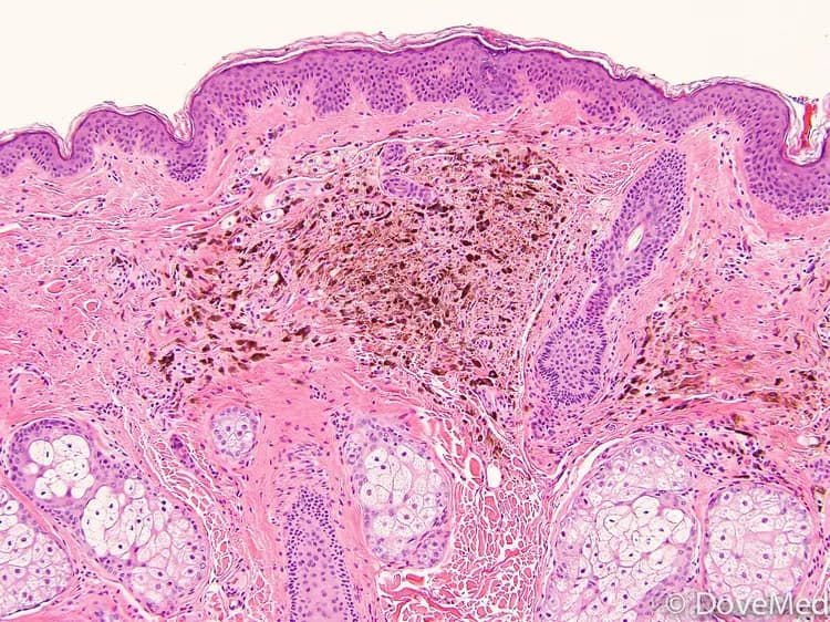
- Skin biopsy: A skin biopsy is performed and sent to a laboratory for a pathological examination. The pathologist examines the biopsy under a microscope. After putting together clinical findings, special studies on tissues (if needed) and with microscope findings, the pathologist arrives at a definitive diagnosis
Many clinical conditions may have similar signs and symptoms. Your healthcare provider may perform additional tests to rule out other clinical conditions to arrive at a definitive diagnosis.
What are the possible Complications of Cellular Blue Nevus?
There are frequently no complications that arise from a Cellular Blue Nevus.
- In some individuals, it may give rise to cosmetic concerns
- In rare cases, a Cellular Blue Nevus can transform into a melanoma (a malignant skin condition). Due to this factor, the nevus is typically removed through surgery
- The uncertainty of a benign or malignant state of the nevus, makes a diagnostic biopsy typically necessary
- Scratching or itching of the moles may lead to bleeding and ulceration. This can cause secondary bacterial or fungal infections to develop
How is Cellular Blue Nevus Treated?
The treatment measures for Cellular Blue Nevus include:
- Surgical excision and removal of the mole
- Blue Nevus can also be excised through electrocautery surgical procedure
- Regular follow up visits may be recommended by the healthcare provider
How can Cellular Blue Nevus be Prevented?
Current medical research has not established a method of preventing the occurrence of Cellular Blue Nevi. However, the following measures can help these moles from either becoming a dysplastic mole or a melanoma:
- Minimizing direct exposure to the sun’s ultraviolet (UV) rays
- Avoiding tanning beds and sun lamps
- Performing self-examination of your skin, from head to toe, once a month (especially if you are at risk)
- Getting a professional skin exam from a healthcare provider, once a year
- Not picking or scratching the moles, which can cause it to ulcerate and bleed
What is the Prognosis of Cellular Blue Nevus? (Outcomes/Resolutions)
- The prognosis of Cellular Blue Nevus is excellent on a complete excision and removal of the mole
- In very rare cases, the nevus may transform into a malignant melanoma; in which case, the prognosis depends upon several factors including the stage of the tumor
Additional and Relevant Useful Information for Cellular Blue Nevus:
The following DoveMed website link is a useful resource for additional information:


Related Articles
Test Your Knowledge
Asked by users
Related Centers
Related Specialties
Related Physicians
Related Procedures
Related Resources
Join DoveHubs
and connect with fellow professionals
0 Comments
Please log in to post a comment.