Background Information:
What are the other Names for the Procedure?
- Pelvic Sonography Procedure
- Pelvic Ultrasound Imaging Procedure
- Ultrasound Imaging of Pelvis
What is Ultrasound Imaging Scan of the Pelvis radiology procedure? (General Explanation)
- Ultrasound imaging is a noninvasive procedure that uses high-frequency sound waves to produce real-time images. The procedure can be used to show movement of internal organs and blood flowing through blood vessels
- An Ultrasound Imaging of Pelvis is used to diagnose various medical conditions in men and women. It includes transabdominal, transvaginal, endovaginal, and transrectal type of imaging
- Doppler ultrasound is useful in assessing blood flow in arteries and veins in arms, legs, abdomen, and neck
What part of the Body does the Procedure involve?
A Pelvic USG Scan images the lower abdomen and genital (pelvic) regions.
Why is Ultrasound Imaging Scan of the Pelvis radiology procedure Performed?
An Ultrasound Imaging of Pelvis is performed to diagnose various medical conditions, which include:
- In women, It is used to evaluate pelvic organs, such as the uterus, cervix, ovaries, and fallopian tubes
- It is also used in pregnancy to evaluate well-being of the fetus and determine age of the pregnancy
- The Ultrasound of Pelvis is used to diagnose pelvic pain, menstrual abnormalities, abnormal bleeding, ovarian cysts, fibroids in uterus, uterine and ovarian cancers
- Transvaginal ultrasounds are used to evaluate the ovaries and lining of uterus (endometrium), which is important in diagnosing uterine anomalies, scars, endometrial polyps, fibroids, and abnormal uterine bleeding
- 3-D ultrasound, also known as sonohysterography, is used to get detailed information about the uterus and irregularities in uterine cavity, in cases of infertility work-up, or to evaluate myometrium (muscular wall of uterus) and endometrium (lining of uterus)
- In men, ultrasound of pelvis is used to evaluate prostate, bladder, and seminal vesicles
- The transrectal ultrasound is specifically designed to evaluate the prostate gland, to a greater detail
- A Pelvic USG Scan is also used to evaluate the kidneys, bladder tumors, and other urinary conditions
What is the Equipment used? (Description of Equipment)
The equipment for ultrasound imaging consists of:
- An ultrasound transducer
- A computer monitor
- A central processing unit
- A printer
A transducer is used to send high frequency sound waves in the body and the computer creates the image based on the echoes of that sound returning from the patient’s body.
What are the Recent Advances in the Procedure?
There have been no recent advances in the field of Pelvic Ultrasound Procedure.
What is the Cost of performing the Ultrasound Imaging Scan of the Pelvis radiology procedure?
The cost of an Ultrasound Imaging of Pelvis procedure depends on a variety of factors, such as the type of your health insurance, annual deductibles, co-pay requirements, out-of-network and in-network of your healthcare providers and healthcare facilities.
In many cases, an estimate may be provided before the procedure. The final amount depends upon the findings during the surgery/procedure and post-operative care that is necessary.
When do you need a Second Opinion, prior to the Procedure?
- It is normal for a patient to feel uncomfortable and confused with a sudden inflow of information regarding the Ultrasound Imaging of Pelvis procedure and what needs to be done
- If the patient needs further reassurance or a second opinion, a physician will almost always assist in recommending another physician
- Also, if the procedure involves multiple steps or has many alternatives, the patient may take a second opinion to understand and choose the best one. They can also choose to approach another physician independently
What are some Helpful Resources?
http://www.radiologyinfo.org/en/info.cfm?pg=genus (accessed on 10/15/2014)
Prior to Ultrasound Imaging Scan of the Pelvis radiology procedure:
How does the Ultrasound Imaging Scan of the Pelvis radiology procedure work?
An Ultrasound Imaging of Pelvis works in the following manner:
- The ultrasound procedure works on similar principles used in sonar by submarines, to determine the size and shape of the object and how far away the object is
- The transducer sends out high-frequency sound waves in the body. When sound waves echo back from the body, the microphone in the transducer records the changes in the sound
- And using the changes in the sound, it is possible to determine the shape, size, or any other changes in the structure of the organ
- Using the change in the echoed sound, a computer produces a picture in real-time on the screen
- Doppler ultrasound is used to determine the direction and speed of the blood cells
How is the Procedure Ultrasound Imaging Scan of the Pelvis radiology procedure Performed?
An Ultrasound Imaging of Pelvis is performed in the following manner:
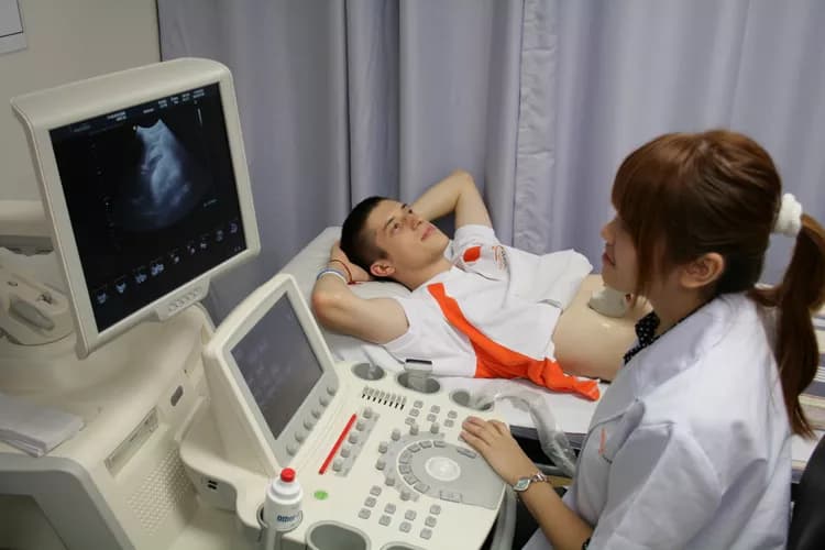
- Transabdominal ultrasound
- The patient lies face-up on the table
- A water-based gel is applied on the area of interest so that transducer can have better contact with the area and air pockets are eliminated between the transducer and the body
- A sonographer puts the transducer on the body and moves it around the area of interest to capture the desired image
- A transvaginal ultrasound involves inserting the transducer into the vagina, to visualize internal organs. The patient is positioned similar to how they are, during a gynecological examination
- A transrectal ultrasound involves inserting the transducer into the patient’s rectum, to visualize the prostate. The patient is made to lie on their sides, with their knees and hips flexed, while facing away from the physician
Where is the Procedure Performed?
The Ultrasound of Pelvis is performed either as an outpatient or inpatient procedure, at a hospital.
Who Performs the Procedure?
An ultrasound technician performs the procedure, under the supervision of a radiologist.
How long will the Procedure take?
The Pelvic Ultrasound Imaging Procedure will take about 30 minutes.
Who interprets the Result?
A radiologist interprets the results of the Ultrasound of Pelvis procedure.
What Preparations are needed, prior to the Procedure?
The following preparations may be needed prior to a Pelvic Ultrasound Imaging Procedure:
- The physician may evaluate the individual’s medical history to gain a comprehensive knowledge of the overall health status of the patient, including information related to the medications that are being currently taken
- Do inform the medical professional if you have a history of any medical conditions, such as a heart disease, asthma, diabetes, or kidney disease
- Do inform the medical professional about any allergies, especially related to barium or iodinated contrast material, which may be used in the procedure
- The ultrasound is not recommended, if the patient had had barium enema or GI tests in the past 2 days
- It is advisable to wear comfortable and loose clothes. Avoid wearing any metal objects or jewelry, as it may interfere with the scan
- It is highly recommended to inform your healthcare professional, if you are pregnant or breastfeeding
- For some specific tests related to the aorta, gallbladder, spleen, liver, or pancreas, the patient may be asked to avoid eating for 8-12 hours before the test
- Depending on the procedure adopted, the patient may be asked for certain bowel or bladder preparations, before the preparation sessions
- For some other tests, the patient may be asked to drink about 6 glasses of water, 2 hours prior to the examination and avoid urinating, in order to keep the bowel full, during the exam
What is the Consent Process before the Procedure?
- A physician will request your consent for the Pelvic Ultrasound Imaging Procedure using an Informed Consent Form
- Consent for the Procedure: A “consent” is your approval to undergo a procedure. A consent form is signed after the risks and benefits of the procedure, and alternative treatment options, are discussed. This process is called informed consent
- You must sign the forms only after you are totally satisfied by the answers to your questions. In case of minors and individuals unable to personally give their consent, the individual’s legal guardian or next of kin, shall give their consent for the procedure
What are the Benefits versus Risks, for this Procedure?
Following are the benefits of the Pelvic Ultrasound Imaging Procedure:
- An Ultrasound of Pelvis provides detailed information about the urinary and reproductive system, to help diagnose various medical conditions
- The ultrasound does not involve the use of any ionizing radiation
- Ultrasound imaging is noninvasive, inexpensive, quick, and simple
- The procedure captures clear images of soft tissues in real-time
- The procedure can be used in needle biopsies or needle aspirations
Following are the risks of Pelvic Ultrasound Imaging Procedure:
- Generally none
What are the Limitations of the Ultrasound Imaging Scan of the Pelvis radiology procedure?
The Pelvic Ultrasound Imaging Procedure cannot be used for examination of the bowel, because air in the bowel disrupts the ultrasound waves.
What are some Questions for your Physician?
Some of the basic questions that you might ask your healthcare provider or physician are as follows:
- What is Ultrasound Imaging of Pelvis procedure?
- Why is this procedure necessary? How will it help?
- How soon should I get it done? Is it an emergency?
- Who are the medical personnel involved in this procedure?
- Where is the procedure performed?
- What are the risks while performing the procedure?
- What are the complications that might take place, during recovery?
- What are the possible side effects from the procedure? How can I minimize these side effects?
- How long will it take to recover? When can I resume normal work?
- How many such procedures have you (the physician) performed?
- Are there any follow-up tests, periodic visits to the healthcare facility required, after the procedure?
- What are the costs involved?
During the Ultrasound Imaging Scan of the Pelvis radiology procedure:
What is to be expected during the Ultrasound Imaging Scan of the Pelvis radiology procedure?
The following may be expected from an USG Pelvic Procedure:
- The test does not cause any pain
- The table you lie on may feel hard, and the room may be cool
- It may be difficult to lie still during the test
- A transvaginal and transrectal ultrasound may cause some discomfort, when the transducer is inserted into the body
- Some individuals may feel nervous during the ultrasound scan
- The healthcare professional will help you during the procedure for optimum images
What kind of Anesthesia is given, during the Procedure?
Anesthesia is rarely used during the Ultrasound Imaging of Pelvis procedure.
How much Blood will you lose, during the Procedure?
Since the procedure is a minimally invasive one, the blood loss involved during the procedure is minimal.
What are the possible Risks and Complications during the Ultrasound Imaging Scan of the Pelvis radiology procedure?
There are no known risks associated with the Ultrasound Imaging of Pelvis procedure.
What Post-Operative Care is needed at the Healthcare Facility after the Ultrasound Imaging Scan of the Pelvis radiology procedure?
No specific post-operative care is needed at healthcare facility after the procedure.
After the Ultrasound Imaging Scan of the Pelvis radiology procedure:
What is to be expected after the Ultrasound Imaging Scan of the Pelvis radiology procedure?
- The Ultrasound Imaging of Pelvis procedure is completely painless, noninvasive, and there is generally no discomfort involved
- However, in the case of the transvaginal and transrectal ultrasound, some discomfort may be felt, when the device is inserted into the body
- The patient may resume their normal activities immediately after the procedure
When do you need to call your Physician?
If the patient is experiencing an allergic reaction from the gel; then, do contact the physician.
What Post-Operative Care is needed at Home after the Ultrasound Imaging Scan of the Pelvis radiology procedure?
No specific post-operative care is needed at home after the Pelvic Ultrasound Imaging Procedure.
How long does it normally take to fully recover, from the Procedure?
The patient needs no recovery time, after the Pelvic Ultrasound Imaging Procedure.
Additional Information:
What happens to tissue (if any), taken out during the Procedure?
- An Ultrasound of Pelvis Procedure does not involve the removal of any body tissue, if the procedure is performed only for imaging of the region
- If a needle biopsy is performed during the scan procedure, a small tissue sample is removed and sent to a pathology laboratory for further evaluation
When should you expect results from the pathologist regarding tissue taken out, during the Procedure?
If a tissue sample is removed during the Ultrasound of Pelvis Procedure and sent for a pathology examination, it may take several days to obtain the results.
Who will you receive a Bill from, after the Ultrasound Imaging Scan of the Pelvis radiology procedure?
It is important to note that the number of bills that the patient may receive depends on the arrangement the healthcare facility has with the physician and other healthcare providers.
Sometimes, the patient may get a single bill that includes the healthcare facility and the consultant physician charges. Sometimes, the patient might get multiple bills depending on the healthcare provider involved. For instance, the patient may get a bill from:
- The hospital, where the procedure is performed
- Healthcare providers, physicians, and radiologists, who are involved in the process
The patient is advised to inquire and confirm the type of billing, before the Ultrasound Imaging of Pelvis procedure is performed.

Related Articles
Test Your Knowledge
Asked by users
Related Centers
Related Specialties
Related Physicians
Related Procedures
Related Resources
Join DoveHubs
and connect with fellow professionals
0 Comments
Please log in to post a comment.