What are the other Names for this Condition? (Also known as/Synonyms)
- SFT (Solitary Fibrous Tumor)
What is Solitary Fibrous Tumor? (Definition/Background Information)
- Solitary Fibrous Tumor (SFT) is a mostly benign (non-cancerous) overgrowth arising from mesenchymal tissue
- The mesenchyme is the middle layer of the 3 primary germ layers of an embryo, namely the ectoderm, mesoderm, and endoderm. The mesoderm gives rise to mesenchymal tissue, which is the source for bone, muscle, connective tissue, and dermis of skin
- Solitary Fibrous Tumors are composed of fibroblasts and related cell types. These rare tumors can occur in any part of the body. The pleura (membrane lining the lung) are the most common site involved
- The cause of formation of the tumor is unknown and currently, no known methods exist to prevent occurrence of the tumor
- Most small tumors are asymptomatic, though the larger ones may compress the surrounding structures and cause related signs and symptoms. The diagnosis of Solitary Fibrous Tumor can be confirmed through a tissue biopsy
- The treatment is complete surgical removal of the tumor with adequate margins. Most Solitary Fibrous Tumors are benign, however, 10-15% of them show malignant behavior in terms of recurrence of the tumor after surgery, or metastasis (spread to distant parts of the body)
- The prognosis of Solitary Fibrous Tumor is dependent on whether the tumor can be completely removed through surgery or not
Solitary Fibrous Tumors may also arise in the soft tissue, or virtually in any part of the body, where mesenchymal cells are present. They have also been reported in the following regions of the body:
- Solitary Fibrous Tumor of Abdominal Wall
- Solitary Fibrous Tumor of Adrenal
- Solitary Fibrous Tumor of Bladder
- Solitary Fibrous Tumor of Breast
- Solitary Fibrous Tumor of Lacrimal Gland
- Solitary Fibrous Tumor of Larynx
- Solitary Fibrous Tumor of Liver
- Solitary Fibrous Tumor of Lung
- Solitary Fibrous Tumor of Major Salivary Glands
- Solitary Fibrous Tumor of Mediastinum
- Solitary Fibrous Tumor of Meninges
- Solitary Fibrous Tumor of Musculoskeletal System, Soft Tissue
- Solitary Fibrous Tumor of Nasal Cavity and Paranasal Sinus
- Solitary Fibrous Tumor of Nasopharynx
- Solitary Fibrous Tumor of Oral Cavity (submucosal mass)
- Solitary Fibrous Tumor of Orbit
- Solitary Fibrous Tumor of Pancreas
- Solitary Fibrous Tumor of Pericardium
- Solitary Fibrous Tumor of Peritoneum (especially pelvis)
- Solitary Fibrous Tumor of Prostate, Seminal Vesicle, and Spermatic Cord
- Solitary Fibrous Tumor of Retroperitoneum
- Solitary Fibrous Tumor of Skin
- Solitary Fibrous Tumor of Pancreas
- Solitary Fibrous Tumor of Thyroid
- Solitary Fibrous Tumor of Ventricles of the Brain
- Solitary Fibrous Tumor of Vulva, Vagina, Cervix, and Endometrium
Who gets Solitary Fibrous Tumor? (Age and Sex Distribution)
- Solitary Fibrous Tumor is a rare tumor with an overall incidence 2.8/100,000. SFTs are rare tumors and account for less than 1% of all soft tissue tumors
- The tumor can be present in individuals between 20 and 70 years of age, but is usually diagnosed in the fifth decade (between 40-50 years)
- In general, both men and women can get it with a similar incidence. However, Solitary Fibrous Tumor of the abdominal wall is more predominant in women
- Though it is a rare tumor, it can occur worldwide and all races and ethnic groups may be affected
What are the Risk Factors for Solitary Fibrous Tumor? (Predisposing Factors)
- No clear-cut risk factors for Solitary Fibrous Tumor have been established to date
It is important to note that having a risk factor does not mean that one will get the condition. A risk factor increases one's chances of getting a condition compared to an individual without the risk factors. Some risk factors are more important than others.
Also, not having a risk factor does not mean that an individual will not get the condition. It is always important to discuss the effect of risk factors with your healthcare provider.
What are the Causes of Solitary Fibrous Tumor? (Etiology)
- Currently, scientists do not know the factor(s) causing Solitary Fibrous Tumor
- Certain genetic mutations are have been detected. Research to characterize these mutations is currently underway
What are the Signs and Symptoms of Solitary Fibrous Tumor?
The signs and symptoms depend on the site of the tumor. They can range in size from a few cm to up to 40 cm; though majority of the tumors are less than 10 cm. They are usually slow-growing and form a single mass. The signs and symptoms associated with Solitary Fibrous Tumor include:
- Small tumors usually do not cause any symptoms. But, occasionally they may become painful, if they compress surrounding structure
- Solitary Fibrous Tumors may occur as slowly enlarging painless mass. The following symptoms may be seen:
- Increased skin temperature over the tumor, if the tumor is visible over the skin
- Telangiectasia (abnormal dilation of red, blue, or purple superficial blood vessels) over the tumor
- Unilateral dilated veins (varicose veins) in the region of the tumor
- Abnormal vibration of pulse (pulsation), or bruit (abnormal sounds heard on auscultation) over the tumor
- Restriction of joint movement if the tumor is close to a joint
- Rarely, Solitary Fibrous Tumors of Soft Tissue, especially if large, may cause hypoglycemia
- Hypoglycemia results in Doege-Potter syndrome, which occurs due to secretion of insulin-like growth factor-2 (IGF-2) by the tumor cells (paraneoplastic syndrome)
- The signs and symptoms of hypoglycemia include confusion, altered mental status, sweating, dizziness, cold hands, etc.
- If the tumor involves the pleura, it is usually asymptomatic. However, sometimes, these tumors can cause symptoms such as cough or shortness of breath. Pleural tumors may have signs and symptoms such as:
- Swelling (clubbing) of fingers or toes
- Joints (also called hypertrophic pulmonary osteoarthropathy)
How is Solitary Fibrous Tumor Diagnosed?
The following procedures and tools may be used in the diagnosis of Solitary Fibrous Tumor:
- Evaluation of the individual’s medical history and a through physical examination
- Plain X-ray of the affected region
- CT or CAT scan with contrast of the affected region usually shows a well-defined mass, which may have calcifications. This radiological procedure creates detailed 3-dimensional images of structures inside the body
- MRI scans of the affected region: Magnetic resonance imaging (MRI) uses a magnetic field to create high-quality pictures of certain parts of the body, such as tissues, muscles, nerves, and bones. These high-quality pictures may reveal the presence of the tumor
- Ultrasound scan of the affected region
- Musculoskeletal Solitary Fibrous Tumor can additionally be evaluated using angiography of the affected region
- MRI scans and PET scans may help differentiate benign versus malignant SFTs by detecting areas of metastasis (if any)
Although the above modalities can be used to make an initial diagnosis, a tissue biopsy of the tumor is necessary to make a definitive diagnosis to begin treatment. The tissue for diagnosis can be procured in multiple different ways which include:
- Fine needle aspiration (FNA) biopsy of the tumor: A FNA biopsy may not be helpful, because one may not be able to visualize the different morphological areas of the tumor. Hence, a FNA biopsy as a diagnostic tool has certain limitations, and an open surgical biopsy is preferred
- Core biopsy of the tumor
- Open biopsy of the tumor
Tissue biopsy:
- A tissue biopsy of the tumor is performed and sent to a laboratory for a pathological examination. A pathologist examines the biopsy under a microscope. After putting together clinical findings, special studies on tissues (if needed) and with microscope findings, the pathologist arrives at a definitive diagnosis. Examination of the biopsy under a microscope by a pathologist is considered to be gold standard in arriving at a conclusive diagnosis
- Biopsy specimens are studied initially using Hematoxylin and Eosin staining. The pathologist then decides on additional studies depending on the clinical situation
- Sometimes, the pathologist may perform special studies, which may include immunohistochemical stains, molecular testing, and very rarely, electron microscopic studies to assist in the diagnosis
Many clinical conditions may have similar signs and symptoms. Your healthcare provider may perform additional tests to rule out other clinical conditions to arrive at a definitive diagnosis.
What are the possible Complications of Solitary Fibrous Tumor?
Complications due to Solitary Fibrous Tumor are dependent upon the location of the tumor and could include:
- Hypoglycemia resulting in Doege-Potter syndrome, which occurs due to secretion of insulin-like growth factor-2 (IGF-2) by the tumor cells
- Recurrence of SFT following surgery
- Malignant transformation resulting in metastatic SFT
- Complications may arise during chemotherapy and radiation therapy, due to the toxic medication or radiation effect
How is Solitary Fibrous Tumor Treated?
There is no standard treatment protocol established for Solitary Fibrous Tumor. However, in majority of cases, a complete surgical excision with clear or wide margins is the preferred mode of treatment, which can result in a cure, especially if it behaves in a benign manner. They are surgically removed with wide margins.
- Long-term follow-up is required, because recurrence at the site of surgery or metastasis in distant sites have been reported many years after surgery, even with tumors that behave benignly
- Radiotherapy can be used as primary therapy in situations where the tumor cannot be removed completely, or when the tumor reappears (recurrent Solitary Fibrous Tumor) after surgery
- Radiotherapy for SFT can also be used as additional therapy after surgery, if there is a possibility of tumor recurrence after surgery, or if there are inadequate margins (possibility of tumor left behind) following surgery. In some cases due to location of tumor, a complete surgical removal of the tumor is difficult
- Chemotherapy can be used for treating Solitary Fibrous Tumor in following conditions:
- When the tumors cannot be removed completely (due to incomplete surgical resection)
- Tumors that recur after surgery (recurrent Solitary Fibrous Tumor)
- Tumors that have spread to distant parts of the body (metastatic Solitary Fibrous Tumor)
- Arterial embolization of Solitary Fibrous Tumor is a possible treatment option. Here the blood supply to the tumor is blocked resulting in tumor death
- Hypoglycemia due to the tumor is treated using corticosteroids
- Post-operative care is important: One must maintain minimum activity levels, until the surgical wound heals
How can Solitary Fibrous Tumor be Prevented?
- Current medical research has not established a way of preventing Solitary Fibrous Tumor formation
- Regular medical screening at periodic intervals with blood tests, radiological scans, and physical examinations, for those who have already endured the tumor, are helpful
What is the Prognosis of Solitary Fibrous Tumor? (Outcomes/Resolutions)
- The most reliable prognostic factor of Solitary Fibrous Tumor is dependent on whether the tumor can be completely removed through surgery with free margins (no traces of the tumor in adjoining tissue) or not
- Current studies show that the tumor does not have any specific histologic feature (when a pathologist examines the tissue under a microscope), which can help in assessing a definite prognosis for the tumor
- Individuals may have an overall survival rate of 54-89% following first complete surgical resection
- Solitary Fibrous Tumor of the pleura, which are attached with a pedicle, or if well-circumscribed have a favorable prognosis
- Up to 12% of Pleural Solitary Fibrous Tumors that behave malignantly can lead to the death of the patient
- As with any tumor, it is important to have follow-up appointments with a physician to monitor for any returning tumors
The prognosis of Solitary Fibrous Tumors also depends upon a set of several factors, which include:
- Stage of tumor: With lower-stage tumors, when the tumor is confined to site of origin, the prognosis is usually excellent with appropriate therapy. In higher-stage tumors, such as tumors with metastasis, the prognosis is poor
- The surgical respectability of the tumor (meaning, if the tumor can be removed completely)
- Overall health of the individual: Individuals with overall excellent health have better prognosis compared with those with poor health
- Age of the individual: Older individuals generally have poorer prognosis than younger individuals
- Whether the tumor is occurring for the first time, or is a recurrent tumor. Recurring tumors have worse prognosis compared to tumors that do not recur
- Response to treatment: Tumors that respond to treatment have better prognosis compared to tumors that do not respond to treatment
An early diagnosis and prompt treatment of the tumor generally yields better outcomes than a late diagnosis and delayed treatment.
Additional and Relevant Useful Information for Solitary Fibrous Tumor:
- A tumor is an abnormal growth of tissue arising due to uncontrolled and rapid multiplication of cells that serve no function. They are also called neoplasms. Tumors can be benign or cancerous. A benign tumor may generally indicate no threat to one’s health; it also means that it is not “cancerous”
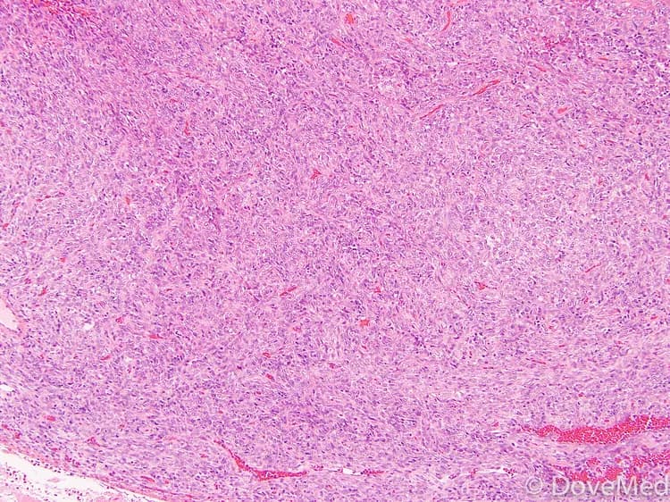
- Grossly, Solitary Fibrous Tumor is well-circumscribed but not encapsulated, and on cross-section appears gray white to yellow white in color
- Microscopically, benign SFT exhibit alternation of hyper- and hypo- cellular areas, patternless pattern, keloid type collagen, and staghorn-shaped vessels-like areas
- A pathologist may use special studies such as special stains. Special stains (markers) used on biopsy samples may include CD34, CD99, Vimentin, BCL-2. These stains help differentiate Solitary Fibrous Tumors from other benign or cancer lesions
- Although Solitary Fibrous Tumor can be classified as malignant based on the biopsy study (tumor greater than 5 cm, increased mitotic rate, necrosis, increased cellularity, or cytologic atypia, infiltrative growth, and weak CD34), these features cannot be absolutely correlated with regards to how it behaves, the nature of which may be aggressive, metastasis, or recurrent
- Basic fibroblast growth factor (bFGF) labeling index and Ki-67 labeling index can be used for evaluation of benign versus malignant Solitary Fibrous Tumors
- Hemangiopericytoma (also known as Solitary Fibrous Tumor - Hemangiopericytoma type; or Cellular Solitary Fibrous Tumor) is closely related to SFT, but is not identical to it. Many pathologists consider hemangiopericytoma and Solitary Fibrous Tumor as a continuum of tumors

Related Articles
Test Your Knowledge
Asked by users
Related Centers
Related Specialties
Related Physicians
Related Procedures
Related Resources
Join DoveHubs
and connect with fellow professionals
0 Comments
Please log in to post a comment.