What are the other Names for this Condition? (Also known as/Synonyms)
- Fibroids of Corpus Uteri
- Leiomyoma of Uterine Corpus
- Uterine Myomas
What is Uterine Fibroids? (Definition/Background Information)
- Uterine Fibroids are the most common tumors of the uterus in young and middle-aged adult women. It is also known as Leiomyoma of Uterine Corpus. A leiomyoma is a benign smooth muscle tumor that can develop anywhere in the body
- Uterine Fibroids are mesenchymal tumors. This indicates that these tumors involve the mesenchyme. The mesenchymal tissue is the source for bone, muscle, connective tissue, and dermis of skin. The fibroids are benign tumors and are not cancers
- It is estimated that a majority of women will have a fibroid during their lifetime. But, these may go unnoticed due to an absence of significant signs and symptoms. In many cases, Uterine Fibroids are diagnosed incidentally, when the women is being examined for other medical conditions
- There are several morphological variants of Leiomyoma of Uterine Corpus (or Uterine Fibroids). These variants include:
- Cellular Leiomyoma of Uterine Corpus
- Leiomyoma of Uterine Corpus with Bizarre Nuclei
- Mitotically-Active Leiomyoma of Uterine Corpus
- Hydropic Leiomyoma of Uterine Corpus
- Leiomyoma of Uterine Corpus with Apoplectic Change
- Lipoleiomyoma (Lipomatous Variant) of Uterine Corpus
- Epithelioid Leiomyoma of Uterine Corpus
- Myxoid Leiomyoma of Uterine Corpus
- Cotyledonoid Dissecting Leiomyoma of Uterine Corpus
- The following are associated conditions of Leiomyoma of Uterine Corpus:
- Intravenous Leiomyomatosis (IVL) of Uterine Corpus
- Diffuse Leiomyomatosis of Uterine Corpus
- Benign Metastasizing Leiomyoma of Uterine Corpus
- Around 90% of the fibroids of the uterus are uncomplicated leiomyomas. The special types or variants (mentioned above) account for about 10% of the uterine leiomyomas
- In many cases, Uterine Fibroids are generally painless and do not show any signs and symptoms. Over a third of the women have pelvic pain and heavy bleeding during menstruation. Large tumors can cause signs and symptoms due to compression of adjoining organ structures
- The treatment of choice for Uterine Fibroids is a surgical removal of the entire tumor. In most cases, the prognosis is excellent with appropriate treatment
Who gets Uterine Fibroids? (Age and Sex Distribution)
- Uterine Fibroids may affect females of any age, but are mostly present in the 30-50 years age category (young and middle-age adults are affected the most)
- These tumors are very common; about 70-80% of women usually have fibroids by the time they are 50 years old
- Women with the rare, genetic disorder, namely hereditary leiomyomatosis and renal cancer syndrome, may present with leiomyomas at an earlier age (while they are much younger)
- Generally, there is no geographical, racial, or ethnic preference noticed
- However, Africans and African Americans have a higher incidence of these tumors, when compared to individuals of other racial groups or ethnicities (3:1 incident rate over other races/ethnic groups). In such individuals, the tumors are often seen to arise at a much younger age
What are the Risk Factors for Uterine Fibroids? (Predisposing Factors)
The risk factors for Uterine Fibroids may include the following:
- Family history of the condition
- Imbalance of estrogen and progesterone hormone levels in the body
- Women of African descent are at a higher risk in comparison to individuals of other races/ethnic groups. In general, such women have larger fibroids, more signs and symptoms, and the tumors are known to grow more rapidly in size
- Early onset of menstruation (in girls)
- Obesity, being overweight
- High in meat and low in vegetables diet
- Vitamin D deficiency
- Excessive alcohol consumption
- Some variants are more common during the active reproductive phase (age) of a woman than others
It is important to note that having a risk factor does not mean that one will get the condition. A risk factor increases ones chances of getting a condition compared to an individual without the risk factors. Some risk factors are more important than others.
Also, not having a risk factor does not mean that an individual will not get the condition. It is always important to discuss the effect of risk factors with your healthcare provider.
What are the Causes of Uterine Fibroids? (Etiology)
- The exact cause of Leiomyoma of Uterine Corpus development is unknown
- In about 40% of the cases, a genetic involvement is noted; chromosomal aberrations or deletions have been observed in chromosomes 6 and 7
- Research is being performed to understand the causative factors of this common condition
What are the Signs and Symptoms of Uterine Fibroids?
A majority of women do not show any signs and symptoms. In others, the signs and symptoms of Uterine Fibroids (or Leiomyoma of Uterine Corpus) typically depend on the tumor location, size, and whether single or multiple tumors are present. The signs and symptom may also depend upon the subtype of leiomyoma.
The following features of Uterine Fibroids may be generally observed in women:
- The leiomyoma tumors are normally multiple in numbers in 3 out of 4 cases
- In some cases, there may be one large tumor with many small-sized tumors or nodules
- The tumors are well-circumscribed and with clear borders
- A wide size range is observed; some are mass-like tumors. The tumors may range in size from a few mm to over 30 cms
- Most tumors are firm, while some are soft; some can be fluid-filled
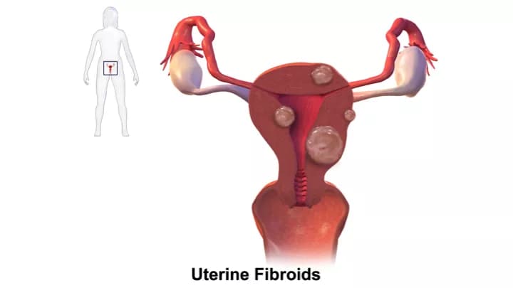
- The tumors may be classified according to the location, and may be:
- Intramural tumors, found within the uterine walls
- Submucosal tumors, found in the thin tissues beneath the mucosal layers
- Subserosal tumors, found in the connective tissues (beneath the serosal layers)
- Tumors at submucosal and subserosal locations are usually like a polyp, or hang in a bag-like structure (known as pedunculated tumors)
The signs and symptoms of Uterine Fibroids may include the following:
- Around 35% of women may have pain in the pelvic region, heavy bleeding during menstruation, and signs and symptoms occurring due to tumor compression
- Women, who are pregnant or under progesterone therapy, may have abdominal pain, discomfort, and other abdomen-related signs and symptoms
- Large tumors may cause a feeling of fullness in the abdomen
- Enlargement of lower abdomen
- Frequent urination due to compression/pressure of the tumor
- Lower back pain
- In a few individuals, the following may be noted:
- Abdominal fluid (ascites)
- Abnormally increased circulating red blood cells, due to the tumor presence (erythropoietin production)
- Presence of leiomyomatosis peritonealis disseminate (a rare condition characterized by multiple smooth muscle tumors)
- Hereditary leiomyomatosis and renal cell cancer (familial form of cancer syndrome showing both uterus and kidney leiomyomas)
- Pain during sexual intercourse
- Bleeding can occur within large tumors (termed as intraleiomyoma hemorrhage)
- Hemorrhage within the tumors can lead to tissue death (or infarction of leiomyoma)
- The bleeding or tissue death within the tumor can also occur during treatment with hormones such as progesterone
How is Uterine Fibroids Diagnosed?
The diagnosis of Uterine Fibroids may involve the following tests and procedures:
- Complete physical examination with thorough evaluation of medical history
- Ultrasound scan of the pelvis: It is a noninvasive procedure that uses high frequency sound waves to produce real-time images
- Abdominal and pelvic CT scan: It is a noninvasive procedure that provides more details of soft tissues, blood vessels, and internal organs
- Pelvic MRI scan: It is a noninvasive medical test that uses a powerful magnetic field to produce images of soft tissues, bones, organs, and all other internal structures of the abdomen and pelvis
- Blood tests that include:
- Complete blood count (CBC) to detect the cause of anemia
- Determination of estrogen/progesterone levels in blood
- Hysteroscopy: This procedure involves placing a probe through the cervix to examine the cavity of the uterus. This exam is helpful in detecting submucosal leiomyomas
- Hysterosalpingography: It is usually performed in individuals with infertility. In this procedure, the structure of the uterus and fallopian tubes are studied by using a dye and X-ray images
- Needle biopsy of tumor: A needle biopsy may not be helpful, because one may not be able to visualize the different morphological areas of the tumor. Hence, a needle biopsy as a diagnostic tool has certain limitations, and an open surgical biopsy is preferred
- Open biopsy of tumor: A tissue biopsy is performed and sent to a laboratory for a pathological examination, who examines the biopsy under a microscope. After putting together clinical findings, special studies on tissues (if needed) and with microscope findings, the pathologist arrives at a definitive diagnosis
- Differential diagnosis, to eliminate other tumor types is considered, before arriving at a definitive diagnosis of Fibroids of Uterine Corpus
Many clinical conditions may have similar signs and symptoms. Your healthcare provider may perform additional tests to rule out other clinical conditions to arrive at a definitive diagnosis.
What are the possible Complications of Uterine Fibroids?
The possible complications of Uterine Fibroids include:
- Stress and anxiety due to fear of cancer of the uterus
- Infertility
- Heavy and prolonged menstrual bleeding may result in anemia
- Polypoid tumors may undergo mechanical injury, such as torsion or twisting, which can result in excruciating pain
- Such polyp-shaped tumors are known to cause cervical prolapse, in some cases
- Fibroids that are attached to the body of the uterus with a pedicle (pedunculated fibroids) may separate from its point of attachment to the body of uterus. Such free-floating fibroids may attach itself to another structure in the abdomen, such as to the pelvic wall, ovary, fallopian tube, or intestines resulting in a parasitic leiomyoma. However, parasitic leiomyomas are not malignant tumors
- Some tumors may grow to large sizes, and even grow out of the uterus affecting adjoining reproductive organs
- Recurrence of the fibroid due to its partial or incomplete surgical removal
- Complications due to underlying disorders such as hereditary leiomyomatosis and renal cell cancer or leiomyomatosis peritonealis disseminate
Complications that may develop in a pregnant woman due to Uterine Fibroids include:
- Pregnancy may cause the fibroids to grow larger in size suddenly
- Loss of pregnancy
- Preterm delivery
- There is a higher incidence of the baby in the womb having an abnormal presentation (such as breech presentation)
- Abruption of placenta: A premature detachment of the placenta from the walls of the uterus, before the baby is delivered
- Intrauterine growth retardation (IUGR): It is a condition when a baby has less than average growth in the womb during pregnancy
- Some study reports indicate that pregnant women with fibroids are 6-times more likely to undergoing a cesarean section (C-section) delivery, than a pregnant woman without the presence of any fibroids
How is Uterine Fibroids Treated?
The treatment of Uterine Fibroids may depend upon a consideration of the following set of factors:
- Size of the fibroid(s)
- Severity of the signs and symptoms
- Location of the fibroids
- Age of the individual
- Whether the women is pregnant or not,
- And the women’s desire to have children in future
The treatment may also depend upon the subtype or variant of the tumor. The following treatment methods may be employed:
- Asymptomatic leiomyomas may not require any treatment; in such cases, the healthcare provider may chose to periodically observe and monitor the tumor
- In some individuals, the size of the fibroids may shrink after menopause without any treatment
- Medical treatment options:
- Pain medications for fibroids causing pain
- Hormonal treatment such as birth control pills
- Dietary and lifestyle modification to address overweight issues
- Taking supplements in case of vitamin D deficiency
- Avoiding alcohol during pregnancy and limiting consumption
- Treating iron-deficiency anemia, if any
- In pregnant women:
- Once the condition is diagnosed, the pregnant mother is closely monitored
- The healthcare provider may recommend an increased frequency of prenatal appointments to monitor progress of the baby’s growth
- Surgical treatment options: A simple surgical excision and removal of the entire tumor is normally sufficient treatment
- Myomectomy: Removal of the fibroids (also known as fibroidectomy)
- Hysterectomy: The removal of a part of the uterus or the entire uterus
- Myolysis of Uterine Fibroids: In this procedure, a needle is inserted into the fibroid. After the insertion, the fibroid is destroyed either by using an electric current, or by a freezing technique
- Uterine Fibroid embolization is a possible treatment option. Here the blood supply to the fibroid is blocked resulting in tumor death
- Radiofrequency ablation: In this technique, the fibroids are destroyed using radio waves
- In case of women with heavy bleeding, endometrial ablation may be performed to control bleeding
- Post-operative care is important: One must maintain minimum activity levels, until the surgical wound heals
- Treatment of any underlying or associated disorder
- Follow-up care with regular screening and check-ups are important
How can Uterine Fibroids be Prevented?
Current medical research has not established a way of preventing Uterine Fibroids, in a majority of women. However, the following factors may be considered to reduce the risk for Uterine Fibroids:
- Address any condition causing hormonal imbalance in the body
- Maintain weight through proper diet modification and physical exercises, if you are overweight/obese
- Avoid alcohol consumption or limit its intake
- Have a balanced diet that is not high in meat and low in vegetables; a balanced diet can also help avoid any mineral or vitamin deficiencies in the body
- Regular prenatal checkups are necessary to monitor the health of the expectant mother and baby in the womb
- Regular medical screening at periodic intervals with blood tests, radiological scans, and physical examinations are mandatory, due to risk of recurrence of the tumor. Often several years of active vigilance is necessary
What is the Prognosis of Uterine Fibroids? (Outcomes/Resolutions)
- In a majority of cases, the prognosis of Uterine Fibroids is generally excellent on surgical excision and removal of the tumor, since these are benign tumors
- The prognosis, in general, depends upon a set of several factors that include:
- The size and number of fibroid(s): Individuals with few (or solitary), small-sized tumors fare better than those with numerous, large-sized tumors
- Overall health of the individual: Individuals with overall excellent health have better prognosis compared with those with poor health
- Age of the individual: Younger women relatively have a better prognosis than older women
- Individuals with less bulky disease have a better prognosis than those with more bulky tumors
- Involvement of vital organs may complicate the condition
- The surgical respectability of the tumor (meaning, if the tumor can be removed completely)
- Whether the tumor is occurring for the first time, or is a recurrent tumor. Recurring tumors have worse prognosis compared to tumors that do not recur
- Response to treatment: Tumors that respond to treatment have better prognosis compared to tumors that do not respond to treatment
- Progression of the condition may make the outcome worse
- An early diagnosis and prompt treatment of the tumor generally yields better outcomes than a late diagnosis and delayed treatment
- Pregnant women with Uterine Fibroids may have a higher risk for complications, which may affect both the mother and baby
- In case of any underlying disorders, the overall prognosis may be dependent upon the severity of the signs and symptoms of the underlying disorder
Additional and Relevant Useful Information for Uterine Fibroids:
- Fibroid tumor removal (or myomectomy) is the surgical removal of fibroids from the uterus
The following link will help you understand fibroid tumor removal surgical procedure:
https://www.dovemed.com/common-procedures/procedures-surgical/fibroid-tumor-removal/
- Hysterectomy is a surgical procedure characterized by the surgical removal of the uterus
The following link will help you understand hysterectomy surgical procedure:
- Uterine fibroid embolization is a minimally-invasive procedure to treat fibroid tumors found in the uterus
The following link will help you understand uterine fibroid embolization radiology procedure:
https://www.dovemed.com/common-procedures/radiology-procedures/uterine-fibroid-embolization/

Related Articles
Test Your Knowledge
Asked by users
Related Centers
Related Specialties
Related Physicians
Related Procedures
Related Resources
Join DoveHubs
and connect with fellow professionals
0 Comments
Please log in to post a comment.