Background Information:
What are the other Names for the Procedure?
- Saline-Infused Sonography
- Sonogram of Uterus
- Ultrasound of Uterus
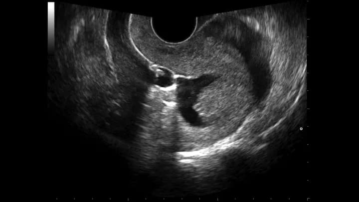
What is Sonohysterography radiology procedure? (General Explanation)
- Sonohysterography is a non-invasive procedure that uses high-frequency sound waves to produce real-time images of the uterus. The procedure can be used to show movement of internal organs and blood flowing through the blood vessels
- Sonohysterography uses ultrasound to provide pictures of the uterus from the inside. It is also known as an Ultrasound of Uterus
- Doppler ultrasound, which evaluates blood flow to the vessel, may also be used as a part of Sonohysterography examination
What part of the Body does the Procedure involve?
The part of the body involved in a Sonohysterography procedure is the uterus.
Why is the Sonohysterography radiology procedure Performed?
Sonohysterography is used for the following purposes:
- To evaluate uterine abnormalities in women with infertility issues or having miscarriages
- To find the cause of unexplained vaginal bleeding in cases of polyps, congenital abnormalities, endometrial adhesions, malignancy, endometrial atrophy, and fibroids
- Doppler ultrasound can also be used in combination with Sonohysterography to determine the blood flow in congenital malformations, AV malformations, aneurysms, polyps, and tumors
What is the Equipment used? (Description of Equipment)
The equipment for Sonohysterography consists of:
- An ultrasound transducer
- A computer monitor
- A central processing unit
- A printer
- A Sonohysterography also uses saline infusion; saline is injected through a thin catheter
A transducer is used to send high-frequency sound waves in the body and the computer creates the image based on the echoes of that sound returning from the patient’s body.
What are the Recent Advances in the Procedure?
There have been no recent advances to the Sonohysterography procedure.
What is the Cost of performing the Sonohysterography radiology procedure?
The cost of the Sonohysterography procedure depends on a variety of factors, such as the type of your health insurance, annual deductibles, co-pay requirements, out-of-network and in-network of your healthcare providers and healthcare facilities.
In many cases, an estimate may be provided before the procedure. The final amount depends upon the findings during the surgery/procedure and post-operative care that is necessary.
When do you need a Second Opinion, prior to the Procedure?
- It is normal for a patient to feel uncomfortable and confused with a sudden inflow of information regarding the Sonohysterography procedure and what needs to be done
- If the patient needs further reassurance or a second opinion, a physician will almost always assist in recommending another physician
- Also, if the procedure involves multiple steps or has many alternatives, the patient may take a second opinion to understand and choose the best one. They can also choose to approach another physician independently
What are some Helpful Resources?
http://www.ncbi.nlm.nih.gov/pubmed/25568988 (assessed on 3/8/2015)
http://www.ncbi.nlm.nih.gov/pubmed/25505226 (assessed on 3/8/2015)
http://www.ncbi.nlm.nih.gov/pubmed/25337156 (assessed on 3/8/2015)
Prior to Sonohysterography radiology procedure:
How does the Sonohysterography radiology procedure work?
A Sonohysterography procedure works in the following manner:
- The ultrasound procedure works on similar principles used in sonar by submarines, to determine the size and shape of the object and how far away the object is
- The transducer sends out high-frequency sound waves in the body. When sound waves echo back from the body, the microphone in the transducer records the changes in the sound
- And using the changes in the sound, it is possible to determine the shape, size, or any other changes in the structure of the organ
- Using the change in the echoed sound, a computer produces a picture in real-time on the screen
- Sonohysterography involves injecting sterile saline into the uterus to distend and enlarge it. Saline outlines the line of uterine cavity (endometrium) and helps visualize polyps or masses within the uterine cavity
- Air can also be used in combination with saline to check the patency of fallopian tube. If air bubbles pass through the fallopian tube, it indicates abnormalities
- Doppler ultrasound is used to determine the direction and speed of the blood cells
How is the Sonohysterography radiology procedure Performed?
A transvaginal ultrasound is usually performed before Sonohysterography, to view the lining of the uterus, its thickness, and any ovarian abnormalities.
- A transvaginal ultrasound involves inserting the transducer into the vagina, to visualize the internal organs. The patient is positioned similar to how they are, during a gynecological examination
- After the transvaginal ultrasound, the probe is removed from the uterus
- A speculum is inserted to open the vagina to visualize the cervix
- The cervix is cleansed and a catheter is inserted into the uterine cavity
- After placement of the catheter, the speculum is removed and an ultrasound probe inserted
- Sterile solution is injected through the catheter into the uterine cavity, as an ultrasound scan is being performed
- After a thorough examination, both catheter and ultrasound probe is removed
Where is the Procedure Performed?
A Sonohysterography is performed as an outpatient procedure, at a hospital.
Who Performs the Procedure?
A radiology technologist performs the Sonohysterography procedure, under the supervision of a gynecologist.
How long will the Procedure take?
- A Sonohysterography procedure takes about 30 minutes to perform
- Sometimes, it may take longer depending upon the complexity of the patient’s anatomy
Who interprets the Result?
A gynecologist interprets the results of the Sonohysterography procedure.
What Preparations are needed, prior to the Procedure?
The following preparations may be needed prior to a Sonohysterography procedure:
- The physician may evaluate the individual’s medical history to gain a comprehensive knowledge of the overall health status of the patient, including information related to the medications that are currently being taken
- Do inform the medical professional if you have a history of any medical conditions such as a heart disease, asthma, diabetes, or kidney disease
- Do inform the medical professional about any allergies, especially related to barium or iodinated contrast material, which may be used in the procedure
- It is advisable to wear comfortable and loose clothes. Avoid wearing any metal objects or jewelry, as it may interfere with the x-ray
- Generally, Sonohysterography is performed one week after menstruation, to avoid any risk of infection. The endometrium is also the thinnest during this period, and thus it is easier to examine the uterus
- Women should notify the physician if they are pregnant or breastfeeding their child, as many such procedures may not be performed on pregnant women
- Depending on the procedure adopted, the patient may be asked for certain bowel or bladder preparations before the preparation sessions
- The patient may be asked to avoid eating or drinking several hours before the test
What is the Consent Process before the Procedure?
A physician will request your consent for the Sonohysterography procedure using an Informed Consent Form.
Consent for the Procedure: A “consent” is your approval to undergo a procedure. A consent form is signed after the risks and benefits of the procedure, and alternative treatment options, are discussed. This process is called informed consent.
You must sign the forms only after you are totally satisfied by the answers to your questions. In case of minors and individuals unable to personally give their consent, the individual’s legal guardian or next of kin, shall give their consent for the procedure
What are the Benefits versus Risks, for this Procedure?
Following are the benefits of Sonohysterography:
- The ultrasound does not involve the use of any ionizing radiation
- Sonohysterography is extremely helpful in visualizing the inside of the uterus and uterine lining
- It provides more details of uterine abnormalities than through a transvaginal ultrasound
- It has much less complications and causes minimal discomfort to the patient
- Ultrasound imaging is noninvasive, inexpensive, quick, and simple
- The procedure captures clear images of the soft tissues in real-time
- The procedure can be used in needle biopsies or needle aspiration
There are no known risks associated with a Sonohysterography radiology procedure.
What are the Limitations of the Sonohysterography radiology procedure?
Following are the limitations of Sonohysterography:
- In certain patients with cervical stenosis, it is difficult to insert a catheter into the uterine cavity for saline infusion
- If the patient has uterine adhesions or fibroids, it becomes extremely difficult to distend the uterine cavity completely, in order to have a good view of the inside of uterus
- It may not be an ideal procedure for determining patency or openness of the fallopian tube. A hysterosalpingography is often performed for those purposes
- Sonohysterography cannot be performed in patients with active pelvic inflammatory disease
What are some Questions for your Physician?
Some of the basic questions that you might ask your healthcare provider or physician are as follows:
- What is a Sonohysterography procedure?
- Why is this procedure necessary? How will it help?
- How soon should I get it done? Is it an emergency?
- Who are the medical personnel involved in this procedure?
- Where is the procedure performed?
- What are the risks while performing the procedure?
- What are the complications that might take place during recovery?
- What are the possible side effects from the procedure? How can I minimize these side effects?
- How long will it take to recover? When can I resume normal work?
- How many such procedures have you (the physician) performed?
- Are there any lifestyle restrictions or modifications required after the procedure is performed?
- Are there any follow-up tests, periodic visits to the healthcare facility required after the procedure?
- Is there any medication that needs to be taken for life after the procedure?
- What are the costs involved?
During the Sonohysterography radiology procedure:
What is to be expected during the Sonohysterography radiology procedure?
- Transvaginal ultrasound and Sonohysterography procedure causes very mild discomfort, but do not cause any pain
- The patient may feel some cramping as the saline is being infused into the uterine cavity to distend it. Over the counter pain medication may be taken before the procedure, to minimize this discomfort
What kind of Anesthesia is given, during the Procedure?
No anesthesia is used during the Sonohysterography procedure.
How much Blood will you lose, during the Procedure?
Since the procedure is a non-invasive one, there is no blood loss involved.
What are the possible Risks and Complications during the Sonohysterography radiology procedure?
There are no risks associated with a Sonohysterography procedure.
What Post-Operative Care is needed at the Healthcare Facility after the Sonohysterography radiology procedure?
No specific post-operative care is needed at healthcare facility after a Sonohysterography procedure.
After the Sonohysterography radiology procedure:
What is to be expected after the Sonohysterography radiology procedure?
- The patient may have vaginal spotting for few days after the Sonohysterography procedure
- They may resume their normal activities immediately after the procedure
When do you need to call your Physician?
Patients may need to call their physician in the following situations after a Sonohysterography procedure:
- If vaginal spotting does not resolve in a few days
- If they develop a high fever
What Post-Operative Care is needed at Home after the Sonohysterography radiology procedure?
No post-operative care is needed at home after the Sonohysterography procedure.
How long does it normally take to fully recover, from the Procedure?
- No recovery time is needed after a Sonohysterography, as it is a noninvasive procedure
- Normal activities may be resumed immediately after the procedure
Additional Information:
What happens to tissue (if any), taken out during the Procedure?
- Generally, the Sonohysterography procedure does not involve the removal of any body tissue
- However, if a biopsy sample is taken during the procedure, it is sent to the pathology lab for further studies
When should you expect results from the pathologist regarding tissue taken out, during the Procedure?
- The tissue removed is processed in the laboratory under a pathologist's supervision
- Slide(s) are prepared once the fluid is processed and is examined by a pathologist and a pathology report issued
- Depending on the complexity of the case, issue of the report may take anywhere between 72 hours to a week's time
Who will you receive a Bill from, after the Sonohysterography radiology procedure?
It is important to note that the number of bills that the patient may receive depends on the arrangement the healthcare facility has with the physician and other healthcare providers.
Sometimes, the patient may get a single bill that includes the healthcare facility and the consultant physician charges. Sometimes, the patient might get multiple bills depending on the healthcare provider involved. For instance, the patient may get a bill from:
- The hospital, where the procedure is performed
- Healthcare providers, physicians, and radiologists, who are involved in the process
The patient is advised to inquire and confirm the type of billing, before the Sonohysterography procedure is performed.

Related Articles
Test Your Knowledge
Asked by users
Related Centers
Related Specialties
Related Physicians
Related Procedures
Related Resources
Join DoveHubs
and connect with fellow professionals
0 Comments
Please log in to post a comment.