What are the other Names for this Condition? (Also known as/Synonyms)
- Adenoma of Sebaceous Gland
- Lipoma of Immature Adipose Tissue
- Pseudolipoma
What is Hibernoma? (Definition/Background Information)
- Hibernoma is a benign tumor of brown fat cells that occurs subcutaneously. They are uncommon and represent about 1% of all tumors of fat
- The most common site of involvement is the thigh. The tumor can also be seen in the head and neck, upper extremity, and trunk (chest and back)
Who gets Hibernoma? (Age and Sex Distribution)
- Hibernomas occurs in young and middle-aged adults (30-40 years age range, with a mean age of 38 years)
- Though it affects both males and females, a slight male predominance is noted
- All racial groups and ethnicities may be affected; no particular predilection is observed
What are the Risk Factors for Hibernoma? (Predisposing Factors)
- There are no known risk factors for Hibernoma at this time
It is important to note that having a risk factor does not mean that one will get the condition. A risk factor increases ones chances of getting a condition compared to an individual without the risk factors. Some risk factors are more important than others.
Also, not having a risk factor does not mean that an individual will not get the condition. It is always important to discuss the effect of risk factors with your healthcare provider.
What are the Causes of Hibernoma? (Etiology)
- Currently, the exact cause of Hibernoma development remains unknown
- However, chromosomal abnormalities in chromosomes 10 and 11 have been associated with this benign tumor
What are the Signs and Symptoms of Hibernoma?
The signs and symptoms of Hibernoma include:
- Hibernoma is an indolent (slow-tumor) tumor producing a mass subcutaneously
- In rare cases, the tumor can occur in a non-subcutaneous location. In a minority of cases (10%), it is found in an intramuscular in location
- These are benign, painless tumors and are usually well-circumscribed, 2-20 cm in size
- It is commonly found in the thigh, followed by the head and neck region, arms, chest and back
How is Hibernoma Diagnosed?
Hibernoma is diagnosed using pathology biopsy and radiology imaging.
- A thorough physical examination and a complete medical history is very important
- Radiographic studies, such as CT or MRI scans of the affected region
- Tissue biopsy: A biopsy is performed and sent to a laboratory for a pathological examination. The pathologist examines the biopsy under a microscope. After putting together clinical findings, special studies on tissues (if needed) and with microscope findings, the pathologist arrives at a definitive diagnosis
Many clinical conditions may have similar signs and symptoms. Your healthcare provider may perform additional tests to rule out other clinical conditions to arrive at a definitive diagnosis.
What are the possible Complications of Hibernoma?
The main complication arising from Hibernoma is that it may keep growing and become a cosmetic issue.
How is Hibernoma Treated?
Hibernomas may be treated through surgery. A surgical excision of the tumor is curative and is adequate.
How can Hibernoma be Prevented?
There are no known preventative measures currently available for Hibernoma.
What is the Prognosis of Hibernoma? (Outcomes/Resolutions)
- Hibernoma is a benign tumor of the fat cells with excellent prognosis on its surgical removal
- They have a benign behavior and there is no metastatic potential
Additional and Relevant Useful Information for Hibernoma:
- Remnant brown fat can be normally seen in the cervical and axillary lymph nodes. In these situations, a diagnosis of Hibernoma should not be made
Following is the diagnostic information on Hibernoma, as observed by a pathologist/radiologist:
Macroscopic (gross) findings: Hibernomas are well-circumscribed tumors that have a lobular cut surface. They are usually large tumors with a median size of 9.3 cm. The cut surface of Hibernoma has fibro-fatty consistency with a yellowish-tan to dark tan appearance.
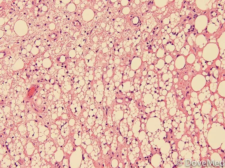
Microscopic (histopathology) findings: Hibernomas show proliferation of brown fat cells which consist of multivacuolated granular cytoplasm with inconspicuous central nucleus. Multivacuolated lipoblast-like cels can be seen which might bring in a differential of well-differentiated liposarcoma occasionally. Myxoid or spindle cells variants have been described. The cytology of Hibernoma cells is bland with no proliferative index.
Special studies - immunohistochemical stains: Hibernomas are positive for S-100, positive for CD34 (spindle cell areas), negative for CD68. Non-spindle cell variants are negative for CD34.
Special studies - genetics and molecular findings: The most common molecular finding is rearrangement of 11Q13. Most of the tumors show numerous chromosomal abnormalities. Homozygous deletion of multiple endocrine neoplasia type I tumor suppress a gene (MEN1) and heterozygous loss of PPP1A have been reported, but is not useful clinically.
Radiology - MRI shows lobular septation which helps in distinguishing it from a lipoma, wherein no lobular septation is visible.
Radiologyy - CT scan shows attenuation of Hibernoma proliferation and shows a contrast between the surrounding fat and skeletal muscle. Hibernomas reveal contrast enhancement.

Related Articles
Test Your Knowledge
Asked by users
Related Centers
Related Specialties
Related Physicians
Related Procedures
Related Resources
Join DoveHubs
and connect with fellow professionals
0 Comments
Please log in to post a comment.